. 2015;59(6):3008-17.
doi: 10.1128/AAC.04830-14. Epub 2015 Mar 9.

Bactericidal efficiency and modes of action of the novel antimicrobial peptide T9W against Pseudomonas aeruginosa

Affiliations

Bactericidal efficiency and modes of action of the novel antimicrobial peptide T9W against Pseudomonas aeruginosa

Xin Zhu et al. Antimicrob Agents Chemother. 2015.

Abstract

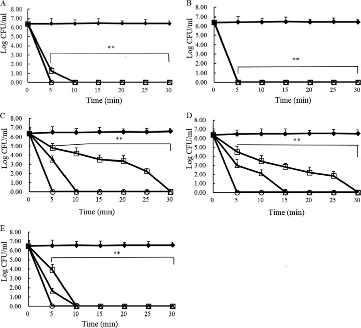
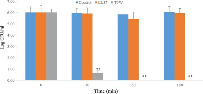
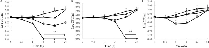
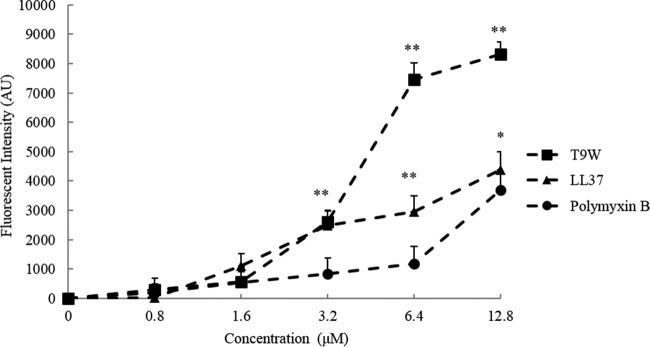
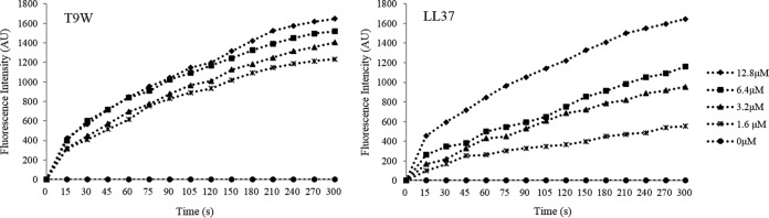
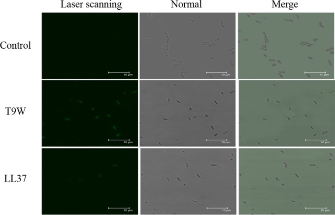
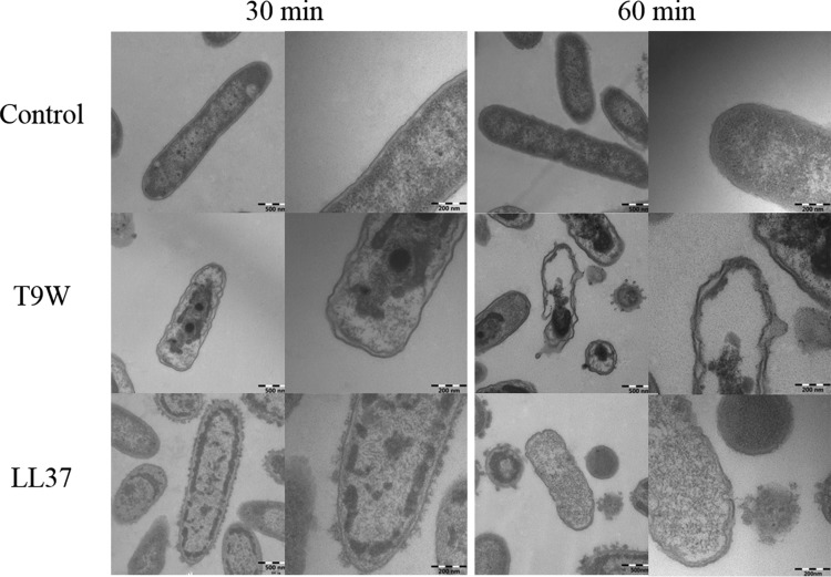
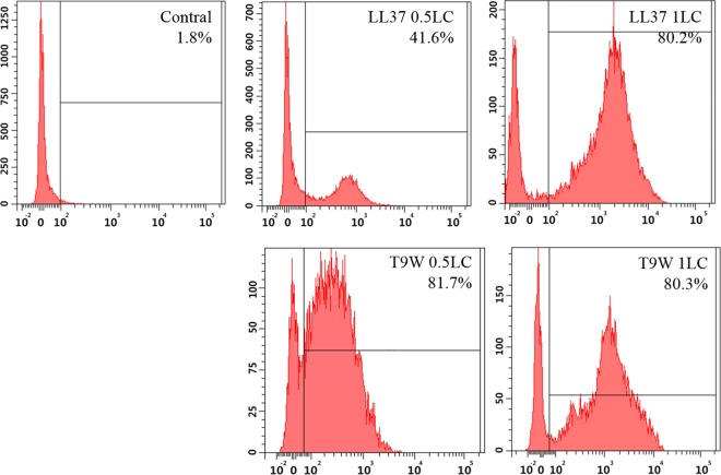
The antipseudomonal efficiency and mechanism of action of a novel engineered antimicrobial peptide, T9W, were evaluated in this study. T9W displayed high activity, with a lethal concentration (LC) of 1 to 4 μM against Pseudomonas aeruginosa, including against ciprofloxacin-, gentamicin-, and ceftazidime-resistant strains, even in the presence of 50 to 300 mM NaCl, 1 to 5 mM Ca(2+), or 0.5 to 2 mM Mg(2+). The time-kill curve (TKC) analysis demonstrated concentration-dependent activity, with T9W achieving complete killing in less than 30 min at 1× LC and in less than 5 min at 4× LC. Combination TKC analyses additionally demonstrated a synergistic effect with ciprofloxacin and gentamicin. The selectivity of T9W was further supported by its ability to specifically eliminate P. aeruginosa in a coculture with macrophages without toxicity to the mammalian cells. The results from fluorescent measurement indicated that T9W bound to lipopolysaccharide (LPS) and induced P. aeruginosa membrane depolarization, and microscopic observations and flow cytometry further indicated that T9W targeted the P. aeruginosa cell membrane and disrupted cytoplasmic membrane integrity, thereby causing cellular content release leading to cell death. This study revealed the potential usefulness of T9W as a novel antimicrobial agent against P. aeruginosa.


Figures

FIG 1
FIG 1
Time-kill kinetics of polymyxin B (A) and T9W (B) against P. aeruginosa 27853 and of T9W against P. aeruginosa LCCI (C), P. aeruginosa LCGE (D), and P. aeruginosa LCCE (E) at 0× LC (filled diamonds), 1× LC (open squares), 2× LC (open triangles), and 4× LC (open circles). Dilutions of aliquots taken from 0 to 30 min were plated on MH agar. The graphs were derived from average values of three independent trials. ∗∗, P < 0.001, compared to the value for the control at the same concentration.
FIG 2
FIG 2
Selective toxicity of T9W in a coculture model. Macrophages (ca. 105 cells/well) were infected with P. aeruginosa 27853 (ca. 107 cells/ml) in RPMI 1640 medium with no bovine fetal serum and antibiotic. The coculture was then treated without or with peptides at 1× LC. The graphs were derived from average values of three independent trials. ∗∗, P < 0.001, compared to the value for the control at the same concentration.
FIG 3
FIG 3
Time-kill kinetics of T9W in combination with ciprofloxacin (A), gentamicin (B), or ceftazidime (C) against P. aeruginosa LCCI (A), P. aeruginosa LCGE (B), or P. aeruginosa LCCE (C). The activity of the combination of 0.5× LC of T9W and 0.5× LC of antibiotic (open diamonds) was compared with the results of 0.5× LC of T9W alone (open squares), 0.5× LC of antibiotic alone (open triangles), or untreated bacteria (open circles). The graphs were derived from average values of three independent trials. ∗, P < 0.01; ∗∗; P < 0.001 (compared to results for the control at the same concentration).
FIG 4
FIG 4
The binding affinity of peptides to the LPS from P. aeruginosa ATCC 27316 (25 μg/ml) was determined by the BODIPY-TR-cadaverine displacement method as described in Materials and Methods. Fluorescence intensity was monitored at an excitation wavelength of 580 nm and an emission wavelength of 620 nm. The graphs were derived from average values of three independent trials. ∗, P < 0.01; ∗∗, P < 0.001 (compared to results with polymyxin B at the same concentration). AU, arbitrary units.
FIG 5
FIG 5
The cytoplasmic membrane potential variation of P. aeruginosa 27853 treated by different concentrations of peptides, as assessed by the release of the membrane potential-sensitive dye diSC3(5). Fluorescence intensity was monitored at an excitation wavelength of 622 nm and an emission wavelength of 670 nm as a function of time. Data plotted are representative average values of three independent experimental trials.
FIG 6
FIG 6
Confocal microscopic images of P. aeruginosa cells. P. aeruginosa 27853 cells were treated with FITC-labeled peptides at 1× LC for 30°C and visualized under laser scanning and transmitted-light scanning (normal) microscopy. Merged profiles are also shown. The control was processed without peptides.
FIG 7
FIG 7
Transmission electron micrographs of P. aeruginosa cells. P. aeruginosa 27853 cells were treated with peptides at 1× LC and visualized at magnifications of ×50,000 (left panels of each pair) and ×120,000 (right panels of each pair). The control was processed without peptides.
FIG 8
FIG 8
Flow cytometric analysis of membrane damage by PI uptake staining. P. aeruginosa 27853 cells were incubated with peptides for 30 min, and the control was processed without peptides.

Similar articles

Cited by

References

    1. Rogers GB, Carroll MP, Bruce KD. 2012. Enhancing the utility of existing antibiotics by targeting bacterial behaviour? Br J Pharmacol 165:845–857. doi:10.1111/j.1476-5381.2011.01643.x. - DOI - PMC - PubMed
    1. Fernandez L, Hancock RE. 2012. Adaptive and mutational resistance: role of porins and efflux pumps in drug resistance. Clin Microbiol Rev 25:661–681. doi:10.1128/CMR.00043-12. - DOI - PMC - PubMed
    1. Fernebro J. 2011. Fighting bacterial infections-future treatment options. Drug Resist Updat 14:125–139. doi:10.1016/j.drup.2011.02.001. - DOI - PubMed
    1. Strateva T, Mitov I. 2011. Contribution of an arsenal of virulence factors to pathogenesis of Pseudomonas aeruginosa infections. Ann Microbiol 61:717–732. doi:10.1007/s13213-011-0273-y. - DOI
    1. Breidenstein EB, de la Fuente-Nunez C, Hancock RE. 2011. Pseudomonas aeruginosa: all roads lead to resistance. Trends Microbiol 19:419–426. doi:10.1016/j.tim.2011.04.005. - DOI - PubMed

Publication types

MeSH terms