. 2015 Mar 31;43(6):3373-88.
doi: 10.1093/nar/gkv173. Epub 2015 Mar 9.

Structural insight into the mechanism of stabilization of the 7SK small nuclear RNA by LARP7

Affiliations

Structural insight into the mechanism of stabilization of the 7SK small nuclear RNA by LARP7

Emiko Uchikawa et al. Nucleic Acids Res. .

Abstract

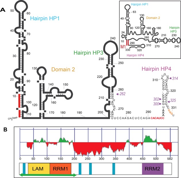
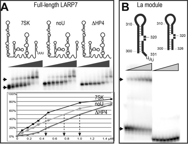
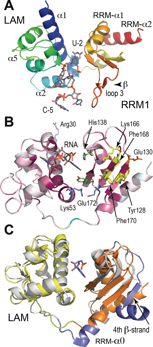
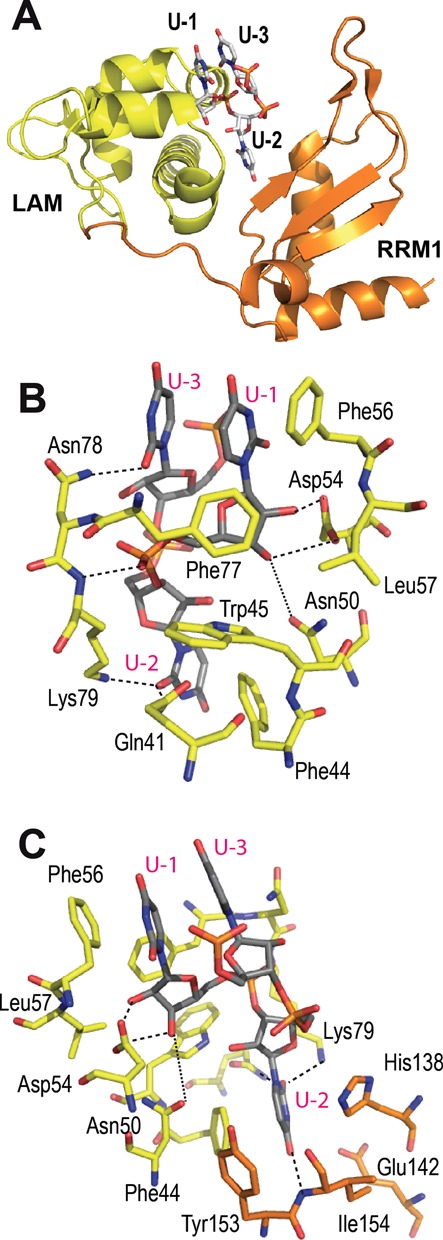
The non-coding RNA 7SK is the scaffold for a small nuclear ribonucleoprotein (7SKsnRNP) which regulates the function of the positive transcription elongation factor P-TEFb in the control of RNA polymerase II elongation in metazoans. The La-related protein LARP7 is a component of the 7SKsnRNP required for stability and function of the RNA. To address the function of LARP7 we determined the crystal structure of its La module, which binds a stretch of uridines at the 3'-end of 7SK. The structure shows that the penultimate uridine is tethered by the two domains, the La-motif and the RNA-recognition motif (RRM1), and reveals that the RRM1 is significantly smaller and more exposed than in the La protein. Sequence analysis suggests that this impacts interaction with 7SK. Binding assays, footprinting and small-angle scattering experiments show that a second RRM domain located at the C-terminus binds the apical loop of the 3' hairpin of 7SK, while the N-terminal domains bind at its foot. Our results suggest that LARP7 uses both its N- and C-terminal domains to stabilize 7SK in a closed structure, which forms by joining conserved sequences at the 5'-end with the foot of the 3' hairpin and has thus functional implications.


Figures

Figure 1.
Figure 1.
Domain organization of the molecules. (A) 2D model of 7SK with the 3′ U-triplet in orange and the additional residue 332 in gray. The two sequences that can form seven base-pairs are in red. The inset shows the closed 2D-structure thus formed. Arrows and numbers in purple indicate the 5′-boundary of the RNAs used in this work, for which the sequence is given. (B) Domain organization of LARP7 (582 amino acids in human) in linear representation with the color scheme adopted for the manuscript: LAM (28–111) yellow, RRM1 (120–188) orange and RRM2 (450–545) purple. The green horizontal arrow corresponds to the protein produced for the structural study. Blue bars represent stretches of basic residues. On top is the prediction of structure formation (red for unfolded, green for folded) as computed with Foldindex.
Figure 2.
Figure 2.
LARP7 N-terminal domain requires the 3′-end uridines for binding. (A) EMSA on a native agarose gel showing complex formation (arrow) after incubation of LARP7 full-length with 7SK: (7SK) wild-type (noU) RNA (1–328) deprived of the terminal U-triplet (ΔHP4) (1–295) deprived of the 3′-hairpin HP4. Complex quantification as a function of protein concentration is reported below. (B) Native gel after incubation with the La module (1–208) showing the free (lower arrow) and the complexed (top arow) RNAs, which are schematized on top: (HP4) hairpin 300–331, (HP4noU) hairpin 300–326 deprived of the terminal U-triplet.
Figure 3.
Figure 3.
Global view of LARP7-Ndom and comparison with La. (A) The La module of LARP7 (LAM domain blue to green, RRM yellow to red) with the RNA in stick representation (gray). (B) Sequence conservation reported in the 3D structure, with ribbon colored from cyan (variable) to wine-red (conserved) and signature residues of RRMs in yellow. LARP7-invariants are highlighted in stick representation: Arg30, Lys32, Lys53, Glu172 (blue), Glu130 (orange), Tyr153, His138 in green. The uridine triplet is shown in gray. (C) Superposition of La (gray) and LARP7 (LAM, yellow and RRM1, orange) with the sequences absent in LARP7 highlighted in blue. U-2, here from LARP7 is at the same position in the two structures.
Figure 4.
Figure 4.
Binding site of the terminal uridines in LARP7. (A) LARP7 La module with LAM domain in yellow and RRM domain in orange, showing the arrangement of the terminal uridines. (B) Details of the 3′ uridines binding site viewed from the LAM side, showing the terminal ribose binding Asn50 and Asp54 and the stacking of U-1 and U-3 with Phe56, in stick representation, with carbons gray for RNA, yellow for amino acids from LAM. Dotted lines indicate H-bonds (distances in the range of 3.0–4.0 Å). (C) Perpendicular view, turned around the vertical axis, showing the specific recognition of U-2 by the RRM1, in orange.
Figure 5.
Figure 5.
The C-terminal domain of LARP7 binds the apical loop of the 3′ hairpin of 7SK. (A) EMSA comparing binding of wild-type RNA HP4 (left panel) and mutant G312C (right panel) with LARP7 full-length (FL), La module (Ndom) or C-terminal domain (Cdom). (B) For each protein, the complex formation with wild-type or mutant HP4 reported as a function of protein concentration. (C) EMSAs of HP4 incubated with La module- (Ndom), C-terminal domain (Cdom) or both domains or mutant G312C (HP4mut) incubated with both domains. The supershift is labeled with a star (*). (D) Binding of the 5′-extended 262-HP4 RNA with the N- (Ndom), C- (Cdom) or both domains, analyzed with a native agarose gel. The supershift is labeled with (*).
Figure 6.
Figure 6.
Localization of the binding sites of the N- and C-terminal domains on the 3′ hairpin of 7SK. (A) Footprinting experiment of the 5′-labeled 262-HP4 RNA with RNases T1 and V1. Denaturing gel showing the cleavage products of the free (R) and complexed RNA with domains N- (N), C- (C), both (NC) or full-length LARP7 (F). Sequence was indexed with T1 in denaturing conditions (G) and ladder (AH); cleavages positions are indicated in green (V1) or blue (T1); control without treatment (C) shows in-line (purple) cleavages. (B) The same experiment but with the mutated 262-HP4 C320G. (C) Summary of the footprinting results. Arrows show cleavages with RNAse T1 (blue), V1 (green) or in-line (pink). Circles represent protections with domains N- (orange) and C-terminal (purple) or full-length LARP7 (or red).
Figure 7.
Figure 7.
Model for the assembly of LARP7 N- and C-terminal domains on the 3′ hairpin of 7SK. (A) Impact of 7SK mutations on the binding with LARP7, estimated by competition experiments. R and X show the migration of the P32-labeled RNA when 7SK is free (R) or bound to full-length LARP7 (X). Increasing amounts (250, 500, 750 or 1000 nM) of mutant RNAs were incubated together with 7SK (50 nM) before adding LARP7. RNAs were 7SK full-length (WT), 1–295 (ΔHP4), 1–328 (noU), mutated at the HP4 bulge (Δ320, U321G) or apical loop (G312C) or at the sequence 289–295 (M1) or deleted of residues 1–8 (Δ9). (B) 2D model showing the M1 region, connecting the 5′-end of 7SK (orange) with the 290–295 sequence (red) and the binding positions of the LARP7 domains N- (orange) and C- (purple) with the linker in green. (C) Our working model, corresponding to the squared region in (B), showing the M1-HP4 RNA with the N-terminal domains (LAM, orange and RRM1, yellow) and the C-terminal domain, represented by the structure of P65 (PDB id. 4ERD, purple). The 5′-terminal phosphate of 7SK is indicated with a green sphere.

Similar articles

Cited by

References

    1. Bousquet-Antonelli C., Deragon J.M. A comprehensive analysis of the La-motif protein superfamily. RNA. 2009;15:750–764. - PMC - PubMed
    1. Maraia R.J., Intine R.V. Recognition of nascent RNA by the human La antigen: conserved and divergent features of structure and function. Mol. Cell. Biol. 2001;21:367–379. - PMC - PubMed
    1. Bayfield M.A., Yang R., Maraia R.J. Conserved and divergent features of the structure and function of La and La-related proteins (LARPs) Biochim. Biophys. Acta. 2010;1799:365–378. - PMC - PubMed
    1. Maris C., Dominguez C., Allain F.H. The RNA recognition motif, a plastic RNA-binding platform to regulate post-transcriptional gene expression. FEBS J. 2005;272:2118–2131. - PubMed
    1. Alfano C., Sanfelice D., Babon J., Kelly G., Jacks A., Curry S., Conte M.R. Structural analysis of cooperative RNA binding by the La motif and central RRM domain of human La protein. Nat. Struct. Mol. Biol. 2004;11:323–329. - PubMed

Publication types

MeSH terms

LinkOut - more resources