. 2015 Mar 31;43(6):3114-27.
doi: 10.1093/nar/gkv181. Epub 2015 Mar 9.

Insulin-response epigenetic activation of Egr-1 and JunB genes at the nuclear periphery by A-type lamin-associated pY19-Caveolin-2 in the inner nuclear membrane

Affiliations

Insulin-response epigenetic activation of Egr-1 and JunB genes at the nuclear periphery by A-type lamin-associated pY19-Caveolin-2 in the inner nuclear membrane

Kyuho Jeong et al. Nucleic Acids Res. .

Abstract

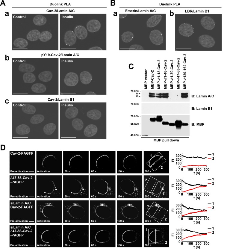
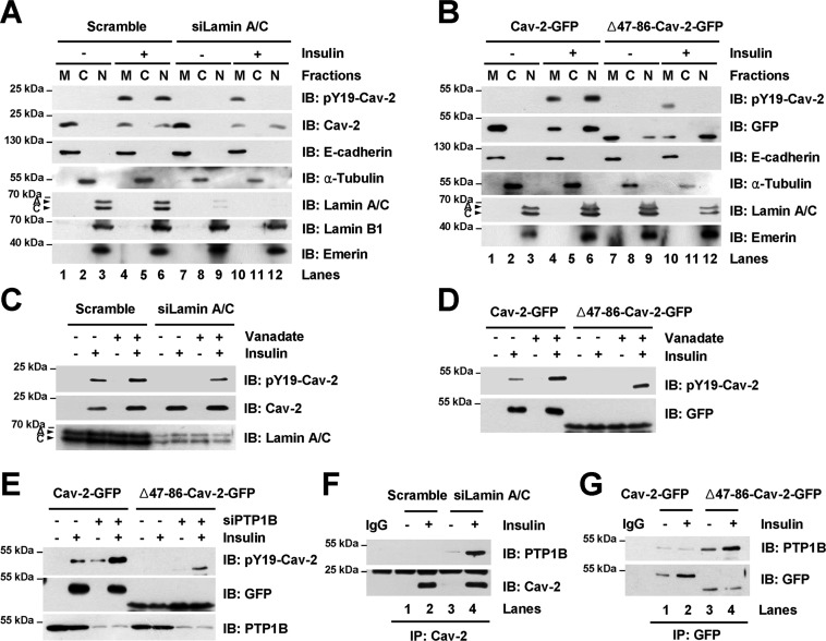
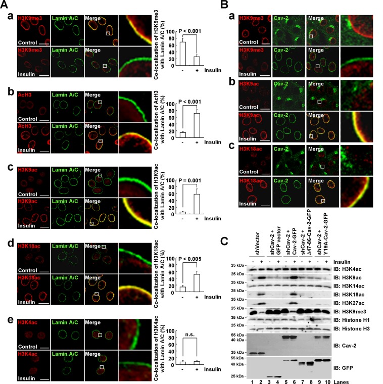
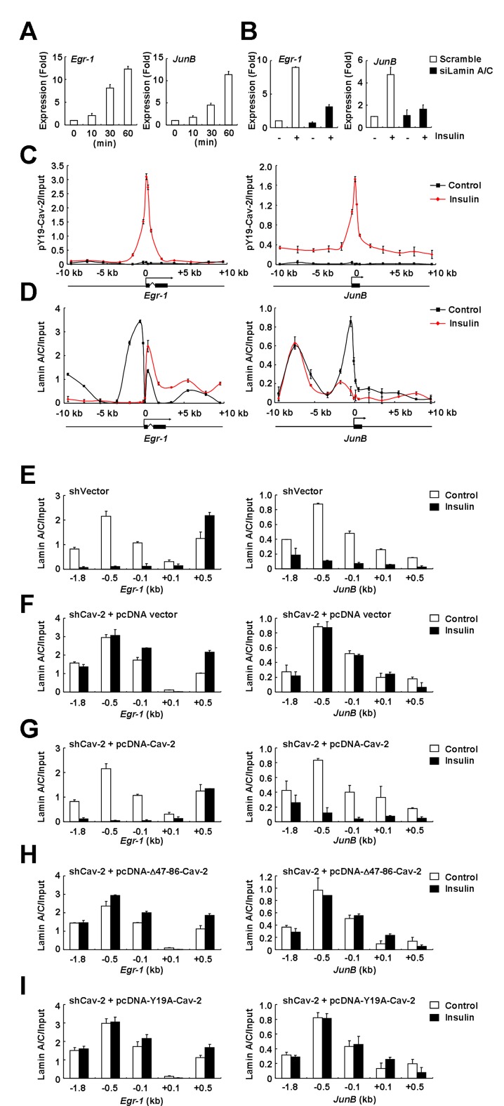
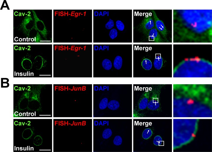
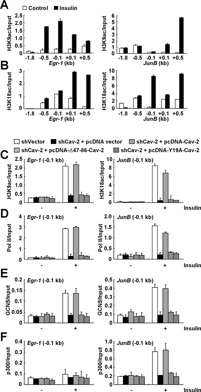
Insulin controls transcription to sustain its physiologic effects for the organism to adapt to environmental changes added to genetic predisposition. Nevertheless, insulin-induced transcriptional regulation by epigenetic factors and in defined nuclear territory remains elusive. Here we show that inner nuclear membrane (INM)-integrated caveolin-2 (Cav-2) regulates insulin-response epigenetic activation of Egr-1 and JunB genes at the nuclear periphery. INM-targeted pY19-Cav-2 in response to insulin associates specifically with the A-type lamin, disengages the repressed Egr-1 and JunB promoters from lamin A/C through disassembly of H3K9me3, and facilitates assembly of H3K9ac, H3K18ac and H3K27ac by recruitment of GCN5 and p300 and the subsequent enrichment of RNA polymerase II (Pol II) on the promoters at the nuclear periphery. Our findings show that Cav-2 is an epigenetic regulator of histone H3 modifications, and provide novel mechanisms of insulin-response epigenetic activation at the nuclear periphery.


Figures

Figure 1.
Figure 1.
pY19-Cav-2 in the INM directly interacts with lamin A/C at the nuclear periphery in response to insulin. (A and B) Cav-2 interaction with lamin A/C but not lamin B1 in endogenous Cav-2-expressing Hirc-B cells. Insulin-induced interaction between Cav-2 and lamin A/C (A, a), pY19-Cav-2 and lamin A/C (A, b), or Cav-2 and lamin B1 (A, c) at the nuclear periphery was determined by Duolink PLA in Hirc-B cells treated with or without 100 nM insulin for 30 min. The in situ interactions between lamin A/C and emerin (B, a) and between lamin B1 and LBR (B, b) in Hirc-B cells were visualized by Duolink PLA. White dots in the gray-scale images represent the clusters of protein–protein interactions. Scale bars, 20 μm. (C) The binding motif of Cav-2 (residues 47–70) to lamin A/C. Nuclear lysates from HEK293T cells expressing no endogenous Caveolins were subjected to in vitro binding assay using MBP vector, MBP-Cav-2, MBP-Δ1–13-Cav-2, MBP-Δ1–46-Cav-2, MBP-Δ1–70-Cav-2, MBP-Δ47–86-Cav-2 or MBP-Δ120–162-Cav-2. (D) Association of Cav-2 in the INM with lamin A/C. HEK293T cells expressed with Cav-2-PAGFP or Δ47–86-Cav-2-PAGFP, or co-transfected with Cav-2-PAGFP or Δ47–86-Cav-2-PAGFP and siLamin A/C were subjected to photoactivation. The graph shows FI of Cav-2-PAGFP or Δ47–86-Cav-2-PAGFP in the NE region before (1: solid box) and after (2: dotted box) photoactivation (mean ± SE, n = 5). Scale bars, 5 μm.
Figure 2.
Figure 2.
pY19-Cav-2 association with lamin A/C is essential for the sustenance of Tyr-19 phosphorylation from the dephosphorylation by nuclear PTP1B. (A and B) Lamin A/C association of Cav-2 in the INM for the maintenance of Tyr-19 phosphorylation. Scramble control or siLamin A/C-transfected Hirc-B cells (A) or Cav-2-GFP- or Δ47–86-Cav-2-GFP-expressed HEK293T cells (B) treated with or without 100 nM insulin for 30 min were subjected to nuclear fractionation. Equal amounts of protein for each fraction were analyzed by immunoblotting (n = 3). E-cadherin, α-tubulin, and lamin A/C and emerin were detected as markers for membrane (M), cytoplasmic (C) and nuclear (N) fractions, respectively. (C and D) Nuclear dephosphorylation of pY19-Cav-2 defective in its association with lamin A/C. Nuclear lysates from scramble control or siLamin A/C-transfected Hirc-B cells (C) or Cav-2-GFP- or Δ47–86-Cav-2-GFP-expressed HEK293T cells (D) treated with or without 200 μM vanadate for 30 min followed by incubation with or without 100 nM insulin for 30 min were subjected to immunoblotting (n = 3). (E) Dephosphorylation of pY19-Cav-2 defective in the lamin A/C association by nuclear PTP1B. Nuclear lysates from Cav-2-GFP or Δ47–86-Cav-2-GFP and scramble control or siPTP1B-co-transfected HEK293T cells treated with or without 100 nM insulin for 30 min were subjected to immunoblotting (n = 3). (F and G) Interaction of the lamin A/C association-defective Cav-2 with nuclear PTP1B. Nuclear lysates from scramble control or siLamin A/C-transfected Hirc-B cells (F) or Cav-2-GFP- or Δ47–86-Cav-2-GFP-expressed HEK293T cells (G) treated with or without 100 nM insulin for 30 min were immunoprecipitated with anti-Cav-2 (F) or anti-GFP (G) antibody and subjected to immunoblotting (n = 3).
Figure 3.
Figure 3.
Lamin A/C-associated pY19-Cav-2 in the INM regulates histone H3 modifications at the nuclear periphery in response to insulin. (A and B) Insulin-induced disassembly of H3K9me3 and assembly of H3K9ac and H3K18ac at the nuclear periphery composed of lamin A/C and Cav-2 in the INM. Hirc-B cells were treated with or without 100 nM insulin for 30 min, stained with anti-H3K9me3 (A, a and B, a), anti-AcH3 (A, b), anti-H3K9ac (A, c and B, b), anti-H3K18ac (A, d and B, c) or anti-H3K4ac (A, e) antibody followed by TRITC-conjugated antibody and with anti-lamin A/C (A) or anti-Cav-2 (B) antibody followed by FITC-conjugated antibody, and analyzed by confocal microscopy (mean ± SE, n = 3). The images on the right show magnifications of the areas framed in the Merge images. Scale bars, 20 μm. n.s., nonsignificant. (C) Regulation of insulin-induced disassembly of H3K9me3 and assembly of H3K9ac, H3K18ac and H3K27ac by lamin A/C-associated pY19-Cav-2. Whole cell lysates from control shRNA vector-expressed Hirc-B cells or GFP vector-expressed Cav-2 shRNA-, Cav-2-GFP-expressed Cav-2 shRNA-, Δ47–86-Cav-2-GFP-expressed Cav-2 shRNA- or Y19A-Cav-2-GFP-expressed Cav-2 shRNA-stable Hirc-B cells treated with or without 100 nM insulin for 30 min were subjected to immunoblotting (n = 4).
Figure 4.
Figure 4.
Lamin A/C-associated pY19-Cav-2 promotes dissociation of the Egr-1 and JunB promoters from lamin A/C in response to insulin. (A) Insulin-stimulated Egr-1 and JunB induction. Hirc-B cells treated with 100 nM insulin for indicated time points were analyzed for Egr-1 and JunB expression by qRT-PCR (mean ± SE, n = 3). (B) Inhibition of insulin-induced Egr-1 and JunB expression by lamin A/C knockdown. Scramble or siLamin A/C-transfected Hirc-B cells treated with or without 100 nM insulin for 30 min were analyzed for Egr-1 and JunB expression by qRT-PCR (mean ± SE, n = 3). (C and D) Insulin-induced enrichment of pY19-Cav-2 around the TSSs (−0.5 to +0.5 kb) and disengagement of the promoters (−0.5 to −0.1 kb) from lamin A/C of Egr-1 and JunB genes. Hirc-B cells treated with or without 100 nM insulin for 30 min were subjected to ChIP with anti-pY19-Cav-2 (C) or anti-lamin A/C (D) antibody on Egr-1 and JunB genes. qPCR data are presented (mean ± SE, n = 3). The intron (zigzag line)/exon (black boxes) organization of the genes is shown at the bottom with an arrow indicating the TSS. (EI) Insulin-induced disengagement of the inactive Egr-1 and JunB promoters from their lamin A/C binding by A-type lamin-associated pY19-Cav-2. Control shRNA vector-expressed Hirc-B cells (E) or pcDNA vector-expressed Cav-2 shRNA- (F), pcDNA-Cav-2-expressed Cav-2 shRNA- (G), pcDNA-Δ47–86-Cav-2-expressed Cav-2 shRNA- (H) or pcDNA-Y19A-Cav-2-expressed Cav-2 shRNA-stable (I) Hirc-B cells treated with or without 100 nM insulin for 30 min were subjected to ChIP with anti-lamin A/C antibody on Egr-1 and JunB genes. qPCR data are presented (mean ± SE, n = 3).
Figure 5.
Figure 5.
Egr-1 and JunB genes are positioned at nuclear periphery and co-localize with the INM-targeted Cav-2 in response to insulin. The in situ localization of Egr-1 and JunB genes at the nuclear periphery was determined by FISH in Hirc-B cells treated with or without 100 nM insulin for 30 min. Representative images of single confocal sections of nuclei show the immunostained Cav-2 (green), FISH signal (red) and DAPI (blue). The arrows in the merge panel indicate the positive signals in the Alexa Fluor 594-labeled Egr-1 (A) and JunB (B) specific DNA probes. The images on the right show magnifications of the areas framed in the merge images. Scale bars, 20 μm.
Figure 6.
Figure 6.
Lamin A/C-associated pY19-Cav-2-mediated recruitment of GCN5 and p300 precedes the subsequent enrichment of H3K9ac and H3K18ac and RNA Pol II on the Egr-1 and JunB promoters in response to insulin. (A and B) Insulin-induced enrichment of H3K9ac and H3K18ac on the Egr-1 and JunB promoters (−0.5 to −0.1 kb). Hirc-B cells treated with or without 100 nM insulin for 30 min were subjected to ChIP with anti-H3K9ac (A) or anti-H3K18ac (B) antibody on Egr-1 and JunB genes. qPCR data in all figures are presented (mean ± SE, n = 4). (C and D) Regulation of the enrichment of H3K9ac on Egr-1 and H3K18ac on JunB promoters and the RNA Pol II recruitment to the promoters (-0.1 kb) by lamin A/C-associated pY19-Cav-2. Control shRNA vector-expressed Hirc-B cells or pcDNA vector-expressed Cav-2 shRNA-, pcDNA-Cav-2-expressed Cav-2 shRNA-, pcDNA-Δ47–86-Cav-2-expressed Cav-2 shRNA- or pcDNA-Y19A-Cav-2-expressed Cav-2 shRNA-stable Hirc-B cells treated with or without 100 nM insulin for 30 min were subjected to ChIP with anti-H3K9ac or anti-H3K18ac antibody (C) or anti-RNA Pol II (D) antibody on the promoters of Egr-1 and JunB genes. qPCR data are presented (mean ± SE, n = 4). (E and F) Recruitment of GCN5 and p300 on the Egr-1 and JunB promoters by lamin A/C-associated pY19-Cav-2. Control shRNA vector-expressed Hirc-B cells or pcDNA vector-expressed Cav-2 shRNA-, pcDNA-Cav-2-expressed Cav-2 shRNA-, pcDNA-Δ47–86-Cav-2-expressed Cav-2 shRNA- or pcDNA-Y19A-Cav-2-expressed Cav-2 shRNA-stable Hirc-B cells treated with or without 100 nM insulin for 30 min were subjected to ChIP with anti-GCN5 (E) or anti-p300 (F) antibody on the promoters of Egr-1 and JunB genes. qPCR data are presented (mean ± SE, n = 4).

Similar articles

Cited by

References

    1. O'Brien R.M., Granner D.K. Regulation of gene expression by insulin. Physiol. Rev. 1996;76:1109–11061. - PubMed
    1. O'Brien R.M., Streeper R.S., Ayala J.E., Stadelmaier B.T., Hornbuckle L.A. Insulin-regulated gene expression. Biochem. Soc. Trans. 2001;29:552–558. - PubMed
    1. Rome S., Clément K., Rabasa-Lhoret R., Loizon E., Poitou C., Barsh G.S., Riou J.P., Laville M., Vidal H. Microarray profiling of human skeletal muscle reveals that insulin regulates approximately 800 genes during a hyperinsulinemic clamp. J. Biol. Chem. 2003;278:18063–18068. - PubMed
    1. Lingwood D., Simons K. Lipid rafts as a membrane-organizing principle. Science. 2010;327:46–50. - PubMed
    1. Parton R.G., del Pozo M.A. Caveolae as plasma membrane sensors, protectors and organizers. Nat. Rev. Mol. Cell Biol. 2013;14:98–112. - PubMed

Publication types

MeSH terms