. 2015 Mar 31;43(6):3046-55.
doi: 10.1093/nar/gkv098. Epub 2015 Mar 9.

Dissecting the behavior and function of MBD3 in DNA methylation homeostasis by single-molecule spectroscopy and microscopy

Affiliations

Dissecting the behavior and function of MBD3 in DNA methylation homeostasis by single-molecule spectroscopy and microscopy

Yi Cui et al. Nucleic Acids Res. .

Abstract

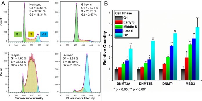
The detailed mechanism for DNA methylation homeostasis relies on an intricate regulatory network with a possible contribution from methyl-CpG-binding domain protein 3 (MBD3). In this study we examine the single-molecule behavior of MBD3 and its functional implication in balancing the activity of DNA methyltransferases (DNMTs). Besides a localization tendency to DNA demethylating sites, MBD3 experiences a concurrent transcription with DNMTs in cell cycle. Fluorescence lifetime correlation spectroscopy (FLCS) and photon counting histogram (PCH) were applied to characterize the chromatin binding kinetics and stoichiometry of MBD3 in different cell phases. In the G1-phase, MBD3, in the context of the Mi-2/NuRD (nucleosome remodeling deacetylase) complex, could adopt a salt-dependent homodimeric association with its target epigenomic loci. Along with cell cycle progression, utilizing fluorescence lifetime imaging microscopy-based Förster resonance energy transfer (FLIM-FRET) we revealed that a proportion of MBD3 and MBD2 would co-localize with DNMT1 during DNA maintenance methylation, providing a proofreading and protective mechanism against a possible excessive methylation by DNMT1. In accordance with our hypothesis, insufficient MBD3 induced by small interfering RNA (siRNA) was found to result in a global DNA hypermethylation as well as increased methylation in the promoter CpG islands (CGIs) of a number of cell cycle related genes.


Figures

Figure 1.
Figure 1.
MBD3 transcription is concurrent with DNMTs during cell cycle progression. (A) Sequential cell synchronization was validated with flow cytometry followed by (B) quantitative RT-PCR for MBD3 and DNMTs. Relative quantity of each gene is normalized to the transcriptional level in the G1-phase and presented as the mean with standard deviation (n = 4). Student's t-test was used to determine the statistical significance.
Figure 2.
Figure 2.
Restriction in the dynamics of nulcear MBD3 increases during cell cycle progression. (A) Illustration of FLCS data collection within different compartments (red cross for nucleus, white cross for cytoplasm) is shown. 2–3 measurement points were randomly chosen from each compartments. (B) Fitting autocorrelation curvers in FLCS: for cytoplamic MBD3-GFP (upper panel) a single-component 3D free diffusion model was used, while for nuclear MBD3-GFP (lower panel) an anomalous diffusion model was applied. (C) The MBD3-GFP diffusion times under different circumstances were obtained. Each group was summarized based on data from more than 15 cells (mean + standard error of the mean, n > 30). The ‘unbound’ measurements were mostly obtained from the free MBD3-GFP in cytoplasm.
Figure 3.
Figure 3.
Analysis of single-molecule brightness by PCH implicates a transition of MBD3 binding stoichiometry. (A) Photon counting histograms from nuclear MBD3-GFP in different cell phases are presented along with corresponding images. (B) Weighted average brightness of different GFP tagged constructs was obtained, with pure GFP as the brightness standard, TLR9-GFP as the dimer control and MBD1-GFP as the monomer control. (C) Dimer percentage for each protein was calculated based on the brightness of pure GFP in buffer. (D) Hypotonic medium disrupted the dimer formation of MBD3-GFP in the G1-phase nucleus. Scale bar: 10 μm.
Figure 4.
Figure 4.
The MBD3–MBD2–DMNT1 complex in DNA maintenance methylation was assessed by FLIM-FRET. (A) Labeled primary antibodies were applied to target the protein pairs (MBD3–DNMT1, MBD3–MBD2). (B) A shortened fluorescence lifetime due to FRET is reflected by the blue-shift in FLIM images (an in vitro antibody-mediated FLIM-FRET is illustrated). (C) FLIM images of the Alexa488 labeled onto anti-MBD3-IgG were obtained in different cell phases, with DNMT1 or MBD2 directly labeled with Alexa555. Scale bar: 10 μm. (D) The ensemble-based fluorescence lifetimes are presented with mean and stand deviation (n = 10 images). FRET efficiencies were accordingly calculated (inset).
Figure 5.
Figure 5.
The dynamic characteristics of MBD3 in DNA methylation homeostasis. In the G1-phase, MBD3/NuRD adopts a ‘two-site sequential binding mode’ on its recognition CGIs and facilitates maintaining a hypomethylated state. kd1 and kd2 denote the two dissociation constants for the homodimeric binding. As the DNA starts to replicate, the newly synthesized hemi-methylated DNA intends to inherit the average methylation density from the parental template, conducted by DNMTs (mainly DNMT1). A proportion of MBD3 and MBD2 (a bona fide 5mC binding protein) would co-operate with DNMT1 herein to complement the accuracy of maintenance methylation. The demethylating potential of MBD3 and MBD2 provides a protective mechanism contributing to DNA methylation homeostasis in the S-G2 phases.
Figure 6.
Figure 6.
Disrupted DNA methylation homeostasis due to insufficient MBD3. (A) The siRNA knockdown effect was verified by quantitative RT-PCR and immunofluorescence. Upon MBD3 knockdown, (B) the global DNA methylation level was quantified with colorimetric immunoassay, (C) and the promoter CGIs methylation of 22 cell cycle related genes was quantified with PCR Array (presented as mean with standard deviation, n = 3). (D) Seventy-two hours after knockdown, the cell cycle distribution was analyzed by flow cytometry.

Similar articles

Cited by

References

    1. Jones P.A. Functions of DNA methylation: islands, start sites, gene bodies and beyond. Nat. Rev. Genet. 2012;13:484–492. - PubMed
    1. Liang G., Chan M.F., Tomigahara Y., Tsai Y.C., Gonzales F.A., Li E., Laird P.W., Jones P.A. Cooperativity between DNA methyltransferases in the maintenance methylation of repetitive elements. Mol. Cell. Biol. 2002;22:480–491. - PMC - PubMed
    1. Lorincz M.C., Schubeler D., Hutchinson S.R., Dickerson D.R., Groudine M. DNA methylation density influences the stability of an epigenetic imprint and Dnmt3a/b—independent de novo methylation. Mol. Cell. Biol. 2002;22:7572–7580. - PMC - PubMed
    1. Walton E.L., Francastel C., Velasco G. Maintenance of DNA methylation: Dnmt3b joins the dance. Epigenetics. 2011;6:1373–1377. - PubMed
    1. Arand J., Spieler D., Karius T., Branco M.R., Meilinger D., Meissner A., Jenuwein T., Xu G., Leonhardt H., Wolf V., et al. In vivo control of CpG and non-CpG DNA methylation by DNA methyltransferases. PLoS Genet. 2012;8:e1002750. - PMC - PubMed

Publication types

MeSH terms

Substances