. 2015 Aug;93(8):891-904.
doi: 10.1007/s00109-015-1264-4. Epub 2015 Mar 11.

Kidney injury is independent of endothelial HIF-1α

Affiliations

Kidney injury is independent of endothelial HIF-1α

Joanna Kalucka et al. J Mol Med (Berl). 2015 Aug.

Abstract

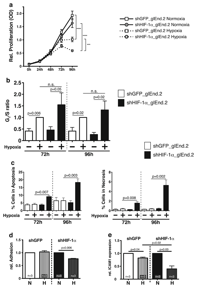
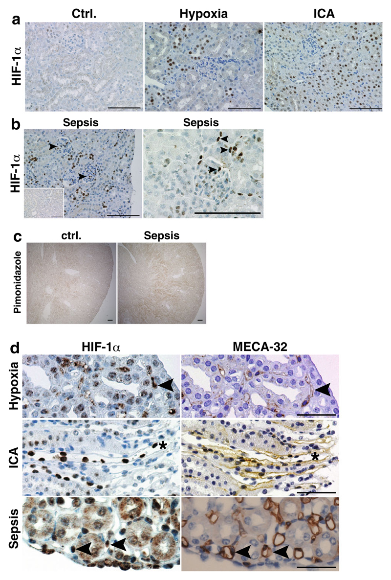
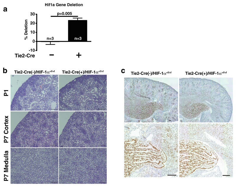
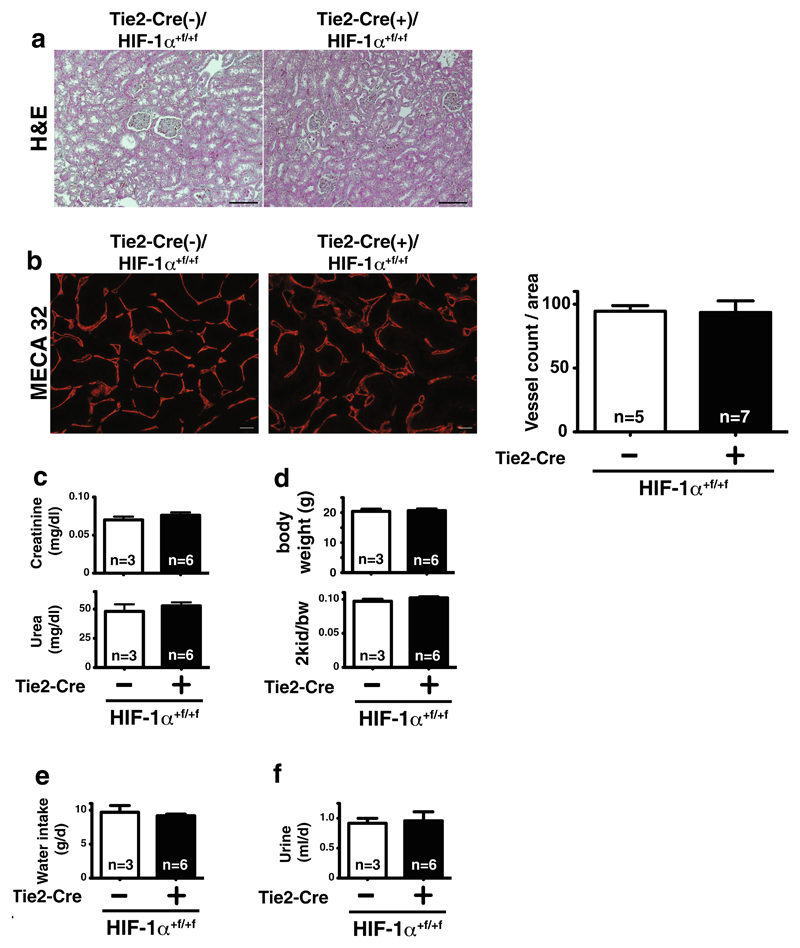
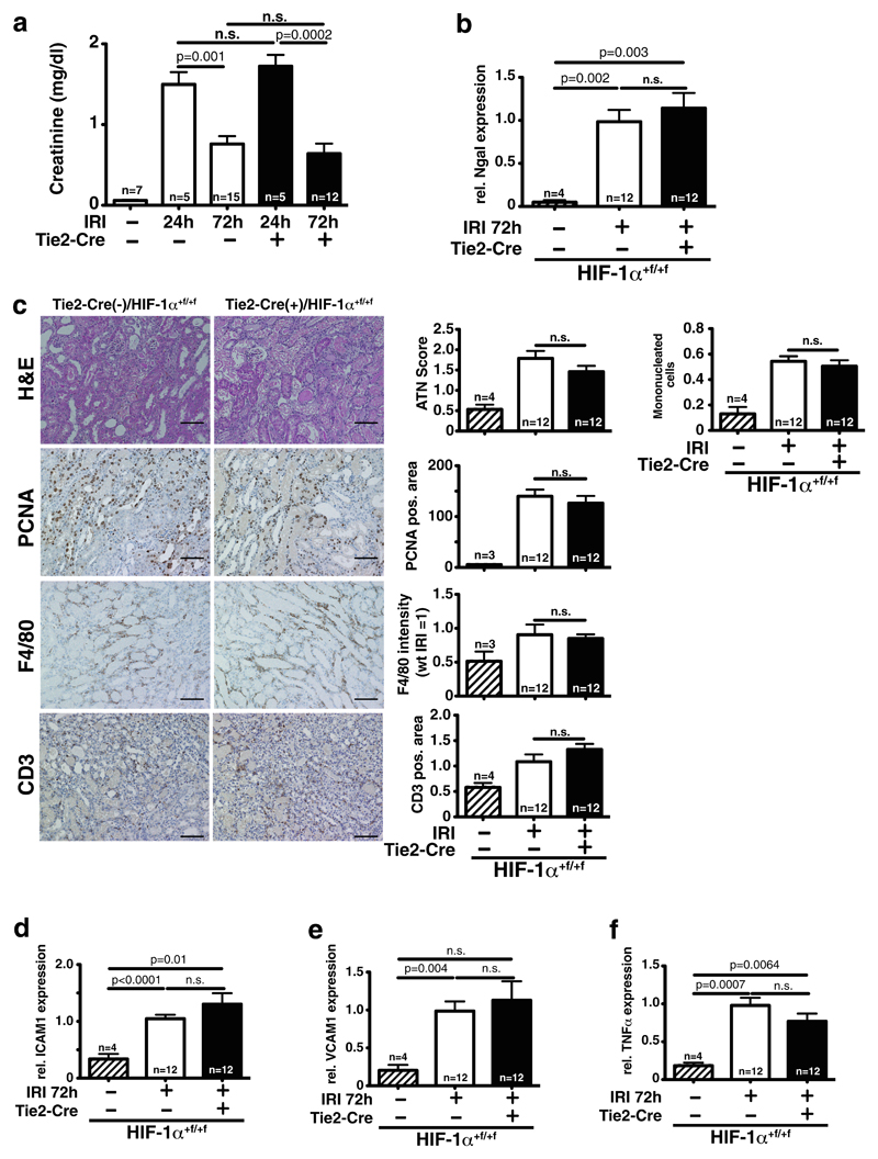
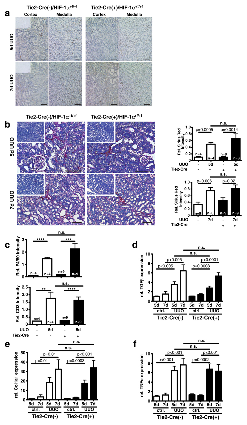
Hypoxia-inducible transcription factors (HIFs) control cellular adaptation to low oxygen. In the kidney, activation of HIF is beneficial during injury; however, the specific contribution of HIF-1α in renal endothelial cells (EC) remains elusive. Since EC display tissue-specific heterogeneity, we investigated how HIF-1α affects key functions of glomerular EC in vitro and its contribution to renal development and pathophysiological adaptation to acute or chronic renal injury in vivo. Loss of HIF-1α in glomerular EC induces hypoxic cell death and reduces hypoxic adhesion of macrophages in vitro. In vivo, HIF-1α expression in EC in mouse kidneys is detectable but limited. Accordingly, EC-specific ablation of HIF-1α does not lead to developmental or phenotypical abnormalities in the kidney. Renal function and expression of adhesion molecules during acute ischemic kidney injury is independent of HIF-1α in EC. Likewise, inflammation and development of fibrosis after unilateral ureteric obstruction is not influenced by endothelial HIF-1α. Taken together, although HIF-1α exerts effects on glomerular EC in vitro, endothelial HIF-1α does not influence renal development and pathophysiological adaptation to kidney injury in vivo. This implies a profound difference of the hypoxic response of the renal vascular bed compared to other organs, such as the heart. This has implications for the development of pharmacological strategies targeting the endothelial hypoxic response pathways.

Key message: HIF-1α controls hypoxic survival and adhesion on endothelial cells (EC) in vitro. In vivo, HIF-1α expression in renal EC is low. Deletion of HIF-1α in EC does not affect kidney development and function in mice. Renal function after acute and chronic kidney injury is independent of HIF-1α in EC. Data suggest organ-specific regulation of HIF-1α function in EC.


Figures

Fig. 1
Fig. 1. Effects of HIF-1α deletion in glomerular microvascular endothelial cells (glEND.2).
a Deletion of HIF-1α in glEND.2 does not affect normoxic proliferation. Hypoxia significantly reduces proliferation starting at 72 h and continuing until 96 h. Deletion of HIF-1α further reduces hypoxic proliferation (n=9–11 per group). b In hypoxic wild-type glEND.2 cells (open bars) G1/S ratio, which indicates cell cycle arrest, is significantly induced compared to normoxia after 72 and 96 h. Deletion of HIF-1α (closed bars) does not significantly change the hypoxic G1/S ratio, thus hypoxic cell cycle arrest is independent of HIF-1α (n >3 per group). c Deletion of HIF-1α (closed bars) significantly induces apoptosis and necrosis in glEnd.2 cells during prolonged hypoxia (n=4–5). d Adhesion of primary macrophages to glEND.2 cells is significantly reduced in hypoxia after deletion of HIF-1α (n =3). e mRNA expression of ICAM1 in glEND.2 cells is significantly reduced after 18 h of hypoxia. Deletion of HIF-1α further reduces hypoxic ICAM1 expression, which indicates that HIF-1α stabilization prevents ICMA1 downregulation in hypoxia (n=3)
Fig. 2
Fig. 2. HIF-1α activation in wild-type kidneys after different stimuli.
a No HIF-1α protein is observed in control conditions. Global HIF-1α activation is more profound after PHD inhibition with ICA than in hypoxia. The majority of HIF-1α protein is detectable in tubular epithelial cells. No glomerular or interstitial HIF-1α staining is visible in hypoxic and ICA-treated kidneys (×400, scale bar 100 μm). b In septic kidneys, tubular, glomerular, and interstitial HIF-1α positivity is observed (arrowheads; ×400, scale bar 100 μm; inset—sham kidney). c Pimonidazole staining confirms hypoxic areas in kidneys of mice treated with CLP-induced sepsis compared to control kidneys (×40, scale bar 100 μm). d Serial sections stained for HIF-1α (left) or the endothelial marker MECA-32 (right) shows specific HIF-1α protein expression (arrowheads) in medullary/papillary EC after stimulation with hypoxia or PHD inhibitors (asterisk denotes the same vessel) or after CLP-induced sepsis (representative images, ×400, scale bar 50 μm)
Fig. 3
Fig. 3. Deletion efficiency, renal development, and HIF-1α protein expression in Tie2-Cre/HIF-1α+f/+f mice.
a Quantification of Hif1a gene in whole kidney DNA shows 23±2.7 % deletion in 9-week-old cre(+) mice (n = 3). b Representative H&E stainings of postnatal kidneys of both genotypes (P1 and P7; ×200, scale bar 100 μm). c Representative HIF-1α staining of P7 postnatal kidneys shows prominent tubular positivity mostly in the papilla in both genotypes (×200, scale bar 100 μm)
Fig. 4
Fig. 4. Renal morphology and functional parameters in adult mice with deletion of HIF-1α in EC.
a H&E-stained kidney sections show no morphological difference in wild-type (Tie2-cre(−)) and endothelial HIF-1α knockout (Tie2-cre(+)) mice (×200, scale bar 100μm). b Immunofluorescent MECA32 stainings (×400) and quantification of renal sections show no differences in renal vessel density (scale bar 20 μm; n=5–7). c Renal function (plasma creatinine (upper panel) and urea (lower panel)) is not significantly different between genotypes. d Body weight (upper panel) and two kidney-to-body weight (bw) ratio (lower panel) as well as water intake (e) and urine production per 24 h (f) in metabolic cages are not affected by deletion of HIF-1α in EC in mice (for all metabolic measurements—WT, n=3; KO, n=6)
Fig. 5
Fig. 5. Loss of endothelial HIF-1α does not affect kidney damage and recovery after ischemia-reperfusion injury (IRI).
a 24 and 72 h after bilateral IRI, renal function of both wild-type and conditional HIF-1α knockout mice is not significantly different (sham, n=7; IRI 24 h, n=5; IRI 72 h, n=12). b 72 h after reperfusion, mRNA of the kidney injury marker Ngal in whole kidney lysates is significantly upregulated compared to sham, but without significant differences between genotypes (sham, n=4; IRI, n=12). c Blinded quantification of tubular injury (ATN score) and infiltrating mononuclear cells on H&E sections shows no difference between wild-type and conditional HIF-1α knockouts 72 h after renal IRI (upper panel). Tubular regeneration (PCNA-positive tubular cells, below) and infiltration of myeloid cells (F4/80 positive) and T cells (CD3 positive) are also not affected by loss of HIF-1α in EC (quantitative analysis of 10 HPF/section; for all histological analyses—sham, n=4; IRI, n=12; shown are representative sections, ×200, scale bar 100 μm). d–f Accordingly, mRNA expression of the adhesion molecules ICAM1 and VCAM1 as well as the proinflammatory cytokine TNFα is significantly upregulated by renal IRI, but deletion of HIF-1α in EC does not change expression (for all mRNA analyses—sham, n=4; IRI, n=12)
Fig. 6
Fig. 6. Loss of endothelial HIF-1α does not affect renal fibrosis in unilateral ureteral obstruction (UUO).
a HIF-1α protein is predominantly activated in tubular cells in the obstructed kidney. Five days after obstruction (top), more HIF-1α expression is visible than after 7 days. No difference in HIF-1α activation is visible between wild-type and endothelial deletion of HIF-1α (representative pictures; ×200, scale bar 100 μm; insets—contralateral control kidney). b Quantification of renal fibrosis by Sirius Red staining with anillin blue counterstain, representative pictures of both genotypes 5 and 7 days after UUO. Insets: representative pictures of contralateral unobstructed kidneys. Five days after UUO (upper panel), fibrotic area increases significantly in both genotypes over the unobstructed kidney without differences after deletion of HIF-1α in EC. Seven days after UUO (lower panel), fibrosis is also significantly increased compared to the unobstructed kidney, without genotype-specific differences (WT, n=4–6; KO, n=8–9; representative pictures; ×200, scale bar 100 μm; insets—contralateral control kidney). c Influx of F4/80-positive cells (upper panel) and CD3-positive T cells (lower panel) is increased after 5 days in the obstructed kidney independent of HIF-1α in EC (WT, n=4; KO, n=9). d Expression of the pro-fibrotic gene TGFβ in total kidney RNA is induced in a time-dependent fashion by UUO, but without influence of endothelial deletion of HIF-1α. e Col1a1 gene expression is also significantly induced in the obstructed kidney in a time-dependent fashion. No genotype-specific difference of expression levels is observed. f mRNA of the proinflammatory cytokine TNFα is induced by UUO independent of HIF-1α in EC (all mRNA analyses—WT, n=4–6; KO, n=9)

Similar articles

Cited by

References

    1. U.S. Renal Data System. USRDS 2013 Annual Data Report: Atlas of End-Stage Renal Disease in the United States. National Institutes of Health, National Institute of Diabetes and Digestive and Kidney Diseases; Bethesda, MD: 2013.
    1. Schurek HJ, Jost U, Baumgartl H, Bertram H, Heckmann U. Evidence for a preglomerular oxygen diffusion shunt in rat renal cortex. Am J Physiol. 1990;259:F910–F915. - PubMed
    1. Fine LG, Norman JT. Chronic hypoxia as a mechanism of progression of chronic kidney diseases: from hypothesis to novel therapeutics. Kidney Int. 2008;74:867–872. - PubMed
    1. Goligorsky MS, Brodsky SV, Noiri E. Nitric oxide in acute renal failure: NOS versus NOS. Kidney Int. 2002;61:855–861. - PubMed
    1. Sun D, Wang Y, Liu C, Zhou X, Li X, Xiao A. Effects of nitric oxide on renal interstitial fibrosis in rats with unilateral ureteral obstruction. Life Sci. 2012;90:900–909. - PubMed

Publication types

MeSH terms

Substances

LinkOut - more resources