. 2015 Jun;14(3):409-20.
doi: 10.1111/acel.12331. Epub 2015 Mar 6.

FSH regulates fat accumulation and redistribution in aging through the Gαi/Ca(2+)/CREB pathway

Affiliations

FSH regulates fat accumulation and redistribution in aging through the Gαi/Ca(2+)/CREB pathway

Xin-Mei Liu et al. Aging Cell. 2015 Jun.

Abstract

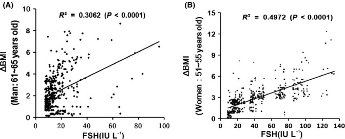
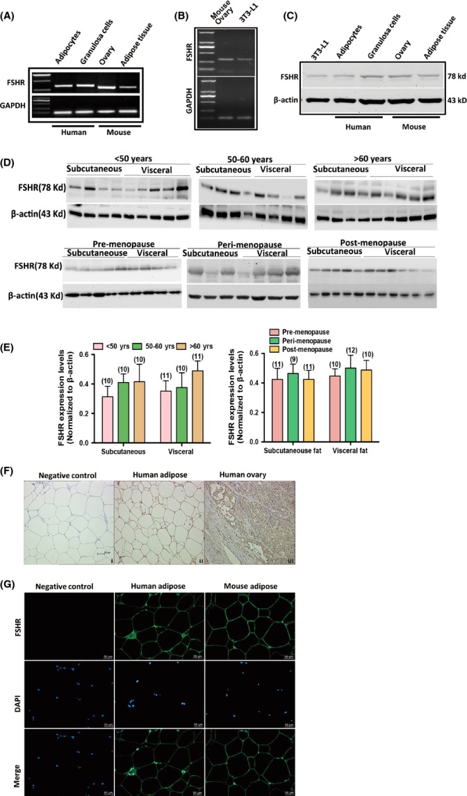
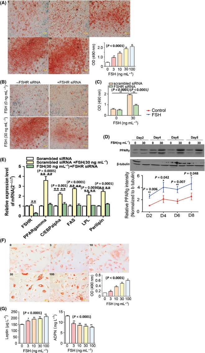
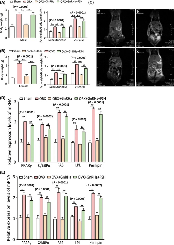
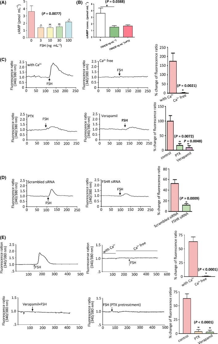
Increased fat mass and fat redistribution are commonly observed in aging populations worldwide. Although decreased circulating levels of sex hormones, androgens and oestrogens have been observed, the exact mechanism of fat accumulation and redistribution during aging remains obscure. In this study, the receptor of follicle-stimulating hormone (FSH), a gonadotropin that increases sharply and persistently with aging in both males and females, is functionally expressed in human and mouse fat tissues and adipocytes. Follicle-stimulating hormone was found to promote lipid biosynthesis and lipid droplet formation; FSH could also alter the secretion of leptin and adiponectin, but not hyperplasia, in vitro and in vivo. The effects of FSH are mediated by FSH receptors coupled to the Gαi protein; as a result, Ca(2+) influx is stimulated, cAMP-response-element-binding protein is phosphorylated, and an array of genes involved in lipid biosynthesis is activated. The present findings depict the potential of FSH receptor-mediated lipodystrophy of adipose tissues in aging. Our results also reveal the mechanism of fat accumulation and redistribution during aging of males and females.

Keywords: ageing; ca2+; endocrinology; mouse models; signal transduction; signalling.


Figures

Fig 1
Fig 1
Correlation between FSH levels and change in BMI (ΔBMI) in aging males and females. (A) Correlation between FSH levels and increase in BMI (ΔBMI) in 414 males (aged 61–65 years). Pearson's = 0.553, < 0.0001. (B) Correlation between FSH levels and increase in BMI (ΔBMI) in 499 females (51–55 years). Pearson's r = 0.710, < 0.0001. ΔBMI(males) = BMI(present) − BMI(aged 35–45 years); ΔBMI(females) = BMI(post-menopausal) − BMI(pre-menopausal).
Fig 2
Fig 2
Expression and localization of FSHR in human and mouse adipocytes. (A) mRNA expressions of FSHR in adipocytes and granulosa cells of human and in adipose tissue and ovarian tissue of mouse. GAPDH served as loading control. (B) mRNA expression of FSHR in 3T3-L1 preadipocytes. GAPDH served as loading control. (C) Protein expressions of FSHR in 3T3-L1 preadipocytes, adipocytes and granulosa cells of human and in adipose tissues and ovarian tissues of mouse. β-actin served as loading control. (D) Protein expression of FSHR in subcutaneous and visceral fat of males aged < 50, 50–60 and > 60 years and in pre-, peri- and postmenopausal females. (E) Relative protein levels of FSHR in subcutaneous and visceral fat of pre-, peri- and postmenopausal females. Values are mean ± SEM; the number of samples in each group is shown at the bottom of the column. No significant differences were observed between groups. (F) Localization of FSHR in human adipose and ovarian tissues by immunohistochemistry. (G) Localization of FSHR in human and mouse adipose tissues by immunofluorescence. Nuclei were stained with DAPI.
Fig 3
Fig 3
FSH-enhanced lipid biosynthesis in 3T3-L1 and human preadipocytes. (A) Concentration-dependent effects of FSH (0–100 ng mL−1) on the differentiation of 3T3-L1 preadipocytes at 8 days. (B, C) Effects of FSHR-targeted siRNA treatment on the differentiation of FSH-induced 3T3-L1 preadipocytes at 8 days. (D) PPARγ expression in FSH-induced 3T3-L1 preadipocytes at 2–8 days of differentiation. (E) PPARγ, C/EBPα, fatty acid synthase, lipoprotein lipase and perilipin transcript levels in FSH-induced 3T3-L1 preadipocytes with or without FSHR-specific siRNA treatment. (F) Concentration-dependent effect of FSH on the differentiation of human preadipocytes at 8 days. (G) Leptin and adiponectin levels in the culture medium of FSH-induced human preadipocyte differentiation at 8 days. Values are mean ± SEM (= 5 for A–G). *< 0.05 and **< 0.01.
Fig 4
Fig 4
Induction of obesity phenotypes and regulation of pro-adipogenic genes by high FSH levels in mice. (A) Body weight variance and subcutaneous and visceral fat mass of male mice in different treatment groups (ORX, orchiectomy). (B) Body weight variance and subcutaneous and visceral fat mass of male mice in different treatment groups (OVX, ovariectomy). (C) Coronal T1-weighted spin-echo MR images obtained with volume segmentation of intra-abdominal tissues from total adipose tissue (a, sham group; b, OVX group; c, OVX+GnRHa group; and d, OVX+GnRHa+FSH group). (D) Transcript levels of lipid biosynthesis-related genes in adipocytes of male mice. (E) Transcript levels of lipid biosynthesis-related genes in adipocytes of female mice. Values are mean ± SEM (= 10 for A and B, = 5 for C–F). < 0.05 and **< 0.01.
Fig 5
Fig 5
Coupling of FSH receptor to Gαi protein and Ca2+ influx in preadipocytes. (A) Intracellular cAMP levels in FSH-induced 3T3-L1 preadipocytes. (B) Intracellular cAMP levels in FSH (30 ng mL−1)-induced 3T3-L1 cells with and without pertussis toxin (PTX). (C) Intracellular Ca2+ measurements in FSH (300 ng mL−1)-treated 3T3-L1 preadipocytes with and without Ca2+ and with PTX (100 nm) and verapamil (20 μm) treatment. (D) Intracellular Ca2+ measurements in FSH (300 ng mL−1)-treated 3T3-L1 preadipocytes with scrambled siRNA and specific FSHR siRNA (7 nm) treatment. (E) Intracellular Ca2+ measurements in FSH-induced human adipocytes with and without Ca2+ and PTX (100 nm) or verapamil (20 μm) treatment. Values are mean ± SEM (= 5 for A–E). *< 0.05, **< 0.01.
Fig 6
Fig 6
Follicle-stimulating hormone-induced pro-lipogenic gene expression via Gαi/Ca2+-dependent CREB/PPARγ pathway in 3T3-L1 preadipocytes. (A) Total CREB and p-CREB expressions in 3T3-L1 cells treated with FSH (0–100 ng mL−1) for 30 min. (B) PPARγ expression in FSH-induced 3T3-L1 cells at 2–8 days of differentiation with or without pertussis toxin (PTX) (100 nm) treatment. (C) FSHR, CREB and p-CREB expressions in 3T3-L1 cells treated with FSH for 30 min with or without FSHR-specific siRNA treatment. (D) CREB and p-CREB expression in 3T3-L1 cells treated with FSH for 30 min with PTX (100 nm) or verapamil (20 μm). (E) Working model of FSH-induced lipid biosynthesis in adipocytes. Values are mean ± SEM (= 5 for A–D). *< 0.05 and **< 0.01.

References

    1. Asatiani K, Gromoll J, Eckardstein SV, Zitzmann M, Nieschlag E, Simoni M. Distribution and function of FSH receptor genetic variants in normal men. Andrologia. 2002;34:172–176. - PubMed
    1. Asayama K, Hayashibe H, Dobashi K, Uchida N, Nakane T, Kodera K, Shirahata A, Taniyama M. Decrease in serum adiponectin level due to obesity and visceral fat accumulation in children. Obes. Res. 2003;11:1072–1079. - PubMed
    1. Ausmanas MK, Tan DA, Jaisamrarn U, Tian XW, Holinka CF. Estradiol, FSH and LH profiles in nine ethnic groups of postmenopausal Asian females: the Pan-Asia Menopause (PAM) study. Climacteric. 2007;10:427–437. - PubMed
    1. Brasaemle DL. Thematic review series: adipocyte biology. The perilipin family of structural lipid droplet proteins: stabilization of lipid droplets and control of lipolysis. J. Lipid Res. 2007;48:2547–2559. - PubMed
    1. Cargill M, Altshuler D, Ireland J, Sklar P, Ardlie K, Patil N, Shaw N, Lane CR, Lim EP, Kalyanaraman N, Nemesh J, Ziaugra L, Friedland L, Rolfe A, Warrington J, Lipshutz R, Daley GQ, Lander ES. Characterization of single-nucleotide polymorphisms in coding regions of human genes. Nat. Genet. 1999;22:231–238. - PubMed

Publication types

Substances