. 2015 Jun;33(6):1850-1862.
doi: 10.1002/stem.1995.

Heparanase released from mesenchymal stem cells activates integrin beta1/HIF-2alpha/Flk-1 signaling and promotes endothelial cell migration and angiogenesis

Affiliations

Heparanase released from mesenchymal stem cells activates integrin beta1/HIF-2alpha/Flk-1 signaling and promotes endothelial cell migration and angiogenesis

Xinyang Hu et al. Stem Cells. 2015 Jun.

Erratum in

  • Corrigendum.
    [No authors listed] [No authors listed] Stem Cells. 2019 May;37(5):E3. doi: 10.1002/stem.3019. Stem Cells. 2019. PMID: 31034132 No abstract available.

Abstract

Heparanase plays important roles in tumor angiogenesis. Our previous study demonstrated that hypoxic preconditioning (HPC) enhanced the angiogenic and therapeutic effects of mesenchymal stem cells (MSCs), effects that were paralleled by enhanced heparanase expression. This study was designed to elucidate the role of heparanase in the improved therapeutic properties of HPC-MSCs and to explore underlying mechanisms using an ischemic rat hind limb model. MSCs transfected with heparanase (MSC(hpa) ) or empty vector (MSC(null) ) were delivered by intramuscular injections to ischemic hind limbs. Hind limbs that received MSC(hpa) recovered blood flow more rapidly at 7 days and acquired higher capillary density at 14 days compared with MSC(null) . Conditioned medium from MSC(hpa) increased endothelial cell migration and promoted greater tube formation relative to that from the MSC(null) groups. Vascular endothelial growth factor receptor 2 (VEGFR2, Flk-1) and its downstream signaling pathway (p38MAPK/HSP27) were significantly increased in human umbilical vein endothelial cells (HUVECs) after treatment with MSC(hpa) conditioned medium. Each of these responses was decreased by cocultured with MSC(hpa-KD) conditioned medium. MSC(hpa) conditioned medium activated hypoxia-inducible factor-2α (HIF-2α) and increased in parallel the transcript level of Flk-1 as determined by chromatin immunoprecipitation-PCR and luciferase assays. Analyses of integrin expression revealed an important role for integrin β1 in the regulation of HIF-2α. All angiogenic effects of MSC(hpa) conditioned medium were abolished by knockdown of integrin β1, HIF-2α, and Flk-1 in HUVECs with selective shRNAs. These findings identify heparanse as a key regulator of angiogenesis by MSCs. We propose a novel pathway wherein heparanse sequentially activates integrin β1, HIF-2α, Flk-1, and p38MAPK/HSP27 with corresponding enhancement of angiogenesis.

Keywords: Angiogenesis; Heparanase; Mesenchymal stem cell.


Figures

Figure 1
Figure 1
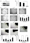
MSChpa promotes vascular regeneration in vivo. (A): Representative Laser-Doppler images (LDI) of hind limbs before, immediately after, and 3, 7, and 14 days after femoral artery occlusion. (B): Quantitative LDI analysis showing the right-to-left (R/L) ratio; n ≥ for each group, * denotes p < .05. (C): Detection of heparanase protein expression using Western blot in WT MSC, MSC harboring empty vector, and heparanase over-expressed vector. (D): Detection of heparanase by ELISA in conditioned medium of WT MSC, MSC harboring empty vector, and heparanase over-expressed vector; n = 4 for each group, * denotes p < .05. (E): HE and immunofluorescent staining of α-SMA and CD31 in muscle tissues from each group; Bar = 100 μm for HE and Bar = 50 μm for immunofluorescent staining. (F, G): Bar graph showed quantitative analysis of SMA and CD31 positive area density; n = 5 for each group, * denotes p < .05; ** denotes p < .01. Abbreviations: MSC, mesenchymal stem cell; PBS, phosphate buffer saline; SMA, smooth muscle actin.
Figure 2
Figure 2
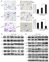
Proangiogenic role of MSChpa in vitro. (A): Protein expression of heparanase and actin by Western blot in MSCnull and MSChpa-KD. (B): Detection of heparanase concentration using ELISA in conditioned medium of MSCWT, MSCnull, and MSChpa-KD; n = 4 for each group, * denotes p < .05. (C): Representative images showing tube formation of human umbilical vein endothelial cells after cocultured with conditioned medium derived from MSCWT, MSCnull, MSChpa, MSChpa-KD, respectively; Bar = 50 μm. (D): Quantification of tube length in each group; n = 3 for each group, ** denotes p < .01. (E): Aortic ring assay showing sprouting and branching in each group; Bar = 50 μm. (F, G): Bar graph showed quantitative analysis of sprout length and outgrowth area, respectively; n = 3 for each group, * denotes p < .05, ** denotes p < .01. Abbreviation: MSC, mesenchymal stem cell.
Figure 3
Figure 3
Enhanced cell migration by MSChpa via Flk-1/p38. (A): Representative images showing cell migration of HUVECs after cocultured with conditioned medium derived from MSCWT, MSCnull, MSChpa, MSChpa-KD, respectively; Bar = 50 μm. (B, C): Bar graph showing quantitative analysis of cell migration of HUVECs in each group; n = 3, * denotes p < .05. (D, E): Quantification of VEGF, Flk-1, Flt-1, Ang1, Tie2, p-p38, p38, p-hsp27, hsp27, p-ERK, ERK, P-AKT, AKT, and actin expression by Western blot in MSCWT, MSCnull, MSChpa, MSChpa-KD, respectively; n = 3. Abbreviations: HUVEC, human umbilical vein endothelial cell; MSC, mesenchymal stem cell.
Figure 4
Figure 4
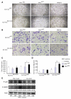
Enhanced HUVEC migration and tube formation is mediated by Flk-1. (A, B): Representative images showing cell migration and tube formation in HUVEC transfected with vector or Flk-1 shRNA lentivirus and cocultured with conditioned medium from MSChpa, MSCnull, DMEM; Bar = 100 μm for tube formation and Bar = 50 μm for migration. (C, D): Bar graph showed quantitative analysis of tube length and cell migration, respectively; n = 3 for each group, * denotes p < .05, ** denotes p < .01. (E): Western blot showing the expression of p-P38, p-hsp27 in HUVEC transfected with vector or Flk-1 shRNA lentivirus; n = 3. Abbreviations: HUVEC, human umbilical vein endothelial cell; MSC, mesenchymal stem cell.
Figure 5
Figure 5
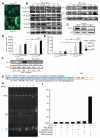
Activation of HIF-2α and Flk-1 by MSChpa. (A): Conditioned medium from MSChpa confers significantly increased nuclear accumulation of HIF-2α in HUVECs, Bar = 25 μm. (B): Quantification of P-NF-KBp105, NF-KBp105, P-NF-KBp50, NF-KBp50, P-NF-KBp65, NF-KBp65, HIF-1α, and HIF-2α expression in HUVEC after coculture with conditioned medium from MSCnull,MSChpa, MSChpa-KD, respectively; n = 3. (C): Quantification of HIF-2α expression in HUVECs transfected with HIF-2α shRNA by Western blot; n = 3 for each group. (D, E): Bar graph showing quantitative analysis of tube length and cell migration, respectively; n = 3 for each group, * denotes p < .05. (F): Western blot showing Flk-1 expression in HUVECs transfected with vector or HIF-2α shRNA lentivirus cultured with conditioned medium from MSCwt, MSCnull, and MSChpa; n = 3. (G, H): Direct binding of HIF-2α to the proximal promoter of Flk-1 confirmed by ChIP-PCR. (I): Luciferase assay showing increased Flk-1 reporter activity by overexpression of HIF-2α. Abbreviations: DMEM, Dulbecco’s modified Eagle’s medium; HUVEC, human umbilical vein endothelial cell; MSC, mesenchymal stem cell.
Figure 6
Figure 6
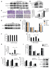
HIF-2α activation by heparanase requires integrin β1. (A): Western blot and quantification of integrin β1 and β3 expressions in HUVECs treated with conditioned medium from MSCnull, MSChpa, and MSChpa-KD. (B): HIF-2α and integrin β1 expressions are decreased in integrin β1 knockdown HUVECs. (C): Representative images and bar graph of cell migration in HUVECs transfected with vector or integrin β1 shRNA lentivirus; Bar = 50 μm, n = 3, ** denotes p < .01. (D): Quantification of VEGF expression in MSCWT, MSCnull, MSChpa-KD, and MSChpa. (E): Quantification of VEGF, PDGF-BB, HGF, and TGF-β concentrations by ELISA in conditioned medium of MSCWT, MSCnull, MSChpa-KD, and MSChpa; n = 4 for each group. * denotes p < .05 (hpa vs. null) and ## denotes p < .01 (hpa-KD vs. null). (F): Viability of MSCs in different concentrations of OGT2115 by CCK-8 assay. (G): Bar graph shows quantitative analysis of cell migration, respectively; n = 3 for each group. * denotes p < .05 (hpa vs. null), ## denotes p < .01 (hpa1OGT2115 vs. hpa). (H): Detection of heparanase concentration by ELISA in conditioned medium of MSCWT, MSCnull, MSChpa, and MSChpa + OGT2115; n = 4 for each group, * denotes p < .05 (hpa vs. null), # denotes p < .05 (hpa+OGT2115 vs. hpa). (I): Detection of VEGF, PDGF-BB, HGF, and TGF-β concentrations by ELISA in conditioned medium of MSChpa and MSChpa with OGT2115, n = 4 for each group. (J, K): Bar graph shows quantitative analysis of cell migration and tube formation in each group; n = 3, ** denotes p < .01, * denotes p < .05. (L): Quantification of integrin β1, Flk-1, and HIF-2α expression in HUVECs after incubated with active heparanase; n = 3. Abbreviations: DMEM, Dulbecco’s modified Eagle’s medium; HUVEC, human umbilical vein endothelial cell; MSC, mesenchymal stem cell.
Figure 7
Figure 7
Schematic of cell migration and angiogenesis by MSC-secreted heparanase. Abbreviation: MSC, mesenchymal stem cell.

References

    1. Heil M, Schaper W. Insights into pathways of arteriogenesis. Curr Pharm Biotechnol. 2007;8:35–42. - PubMed
    1. Koneru S, Varma Penumathsa S, Thirunavukkarasu M, et al. Sildenafil-mediated neovascularization and protection against myocardial ischaemia reperfusion injury in rats: Role of VEGF/angiopoietin-1. J Cell Mol Med. 2008;12:2651–2664. - PMC - PubMed
    1. Liang H, Hou H, Yi W, et al. Increased expression of pigment epithelium-derived factor in aged mesenchymal stem cells impairs their therapeutic efficacy for attenuating myocardial infarction injury. Eur Heart J. 2013;34:1681–1690. - PMC - PubMed
    1. Poitevin S, Cussac D, Leroyer AS, et al. Sphingosine kinase 1 expressed by endothelial colony-forming cells has a critical role in their revascularization activity. Cardiovasc Res. 2014;103:121–130. - PubMed
    1. Chamberlain G, Fox J, Ashton B, et al. Concise review: Mesenchymal stem cells: Their phenotype, differentiation capacity, immunological features, and potential for homing. Stem Cells. 2007;25:2739–2749. - PubMed

Publication types

MeSH terms

Substances