. 2015 Mar 19;22(3):355-68.
doi: 10.1016/j.chembiol.2015.02.003. Epub 2015 Mar 5.

Activation of muscular TrkB by its small molecular agonist 7,8-dihydroxyflavone sex-dependently regulates energy metabolism in diet-induced obese mice

Affiliations

Activation of muscular TrkB by its small molecular agonist 7,8-dihydroxyflavone sex-dependently regulates energy metabolism in diet-induced obese mice

Chi Bun Chan et al. Chem Biol. .

Abstract

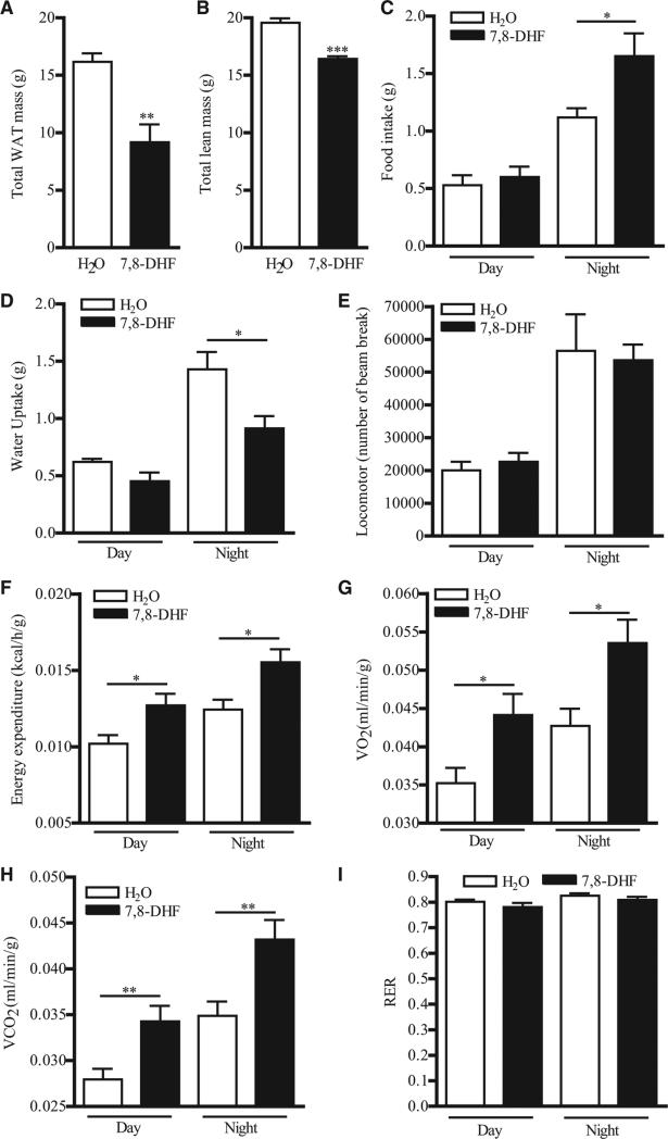
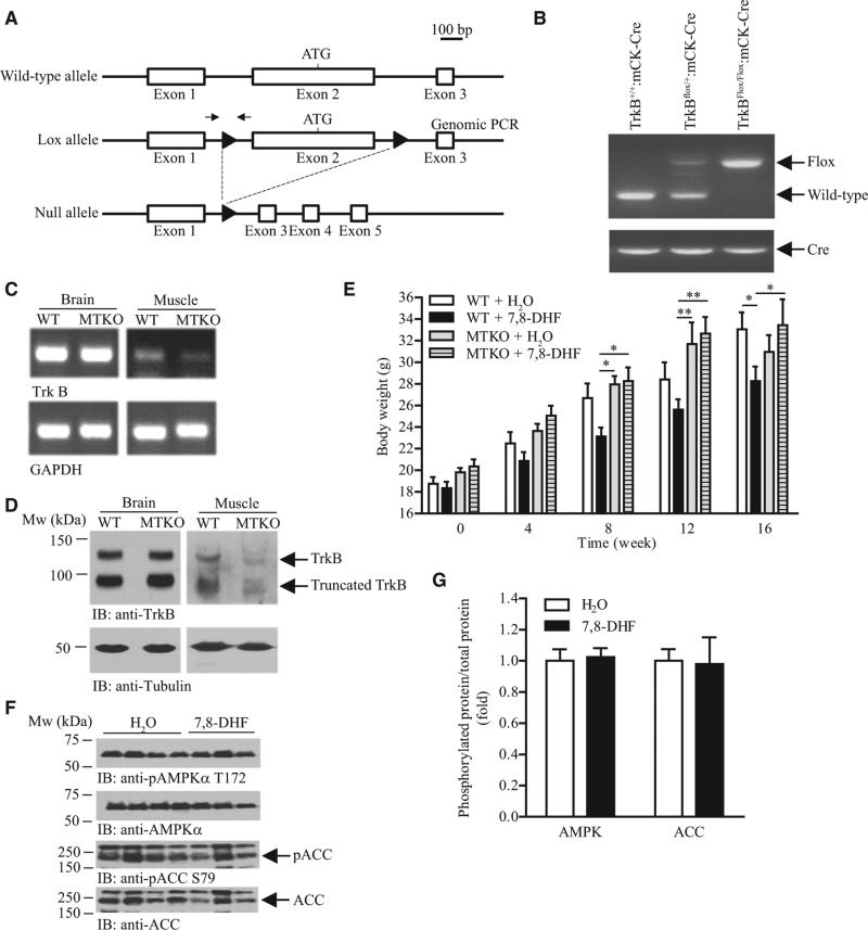
Chronic activation of brain-derived neurotrophic factor (BDNF) receptor TrkB is a potential method to prevent development of obesity, but the short half-life and nonbioavailable nature of BDNF hampers validation of the hypothesis. We report here that activation of muscular TrkB by the BDNF mimetic, 7,8-dihydroxyflavone (7,8-DHF), is sufficient to protect the development of diet-induced obesity in female mice. Using in vitro and in vivo models, we found that 7,8-DHF treatment enhanced the expression of uncoupling protein 1 (UCP1) and AMP-activated protein kinase (AMPK) activity in skeletal muscle, which resulted in increased systemic energy expenditure, reduced adiposity, and improved insulin sensitivity in female mice fed a high-fat diet. This antiobesity activity of 7,8-DHF is muscular TrkB-dependent as 7,8-DHF cannot mitigate diet-induced obesity in female muscle-specific TrkB knockout mice. Hence, our data reveal that chronic activation of muscular TrkB is useful in alleviating obesity and its complications.


Figures

Figure 1
Figure 1. 7,8-DHF-Treated Female Mice Are Resistant to Diet-Induced Obesity
(A) Growth curve of 8-week-old female mice fed with different combinations of diets (**P < 0.01, **P < 0.001, two-way ANOVA versus the same diet feeding, n = 8–10). (B) Organ weight of female mice that have been fed with HFD and 7,8-DHF for 20 weeks (**P < 0.01, Student's t test, n = 5). (C) Pictures of H&E staining of inguinal WAT and liver sections from female mice that have been fed with HFD and 7,8-DHF for 20 weeks. Representative results of four different mice from each treatment are shown. Scale bar represents 50 mm. (D) Adipocyte size of inguinal fat pad isolated from female mice that have been fed with HFD and 7,8-DHF for 20 weeks (n = 4). (E) Circulating leptin concentration in female mice that have been fed with HFD and 7,8-DHF for 20 weeks (*P < 0.05, Student's t test, n = 6). (F) Circulating TNFα concentration in female mice that have been fed with HFD and 7,8-DHF for 20 weeks (*P < 0.05, Student's t test, n = 6). (G) Tissue cholesterol content in female mice that have been fed with HFD and 7,8-DHF for 20 weeks (*P < 0.05, Student's t test, n = 6). (H) Tissue TG content in female mice that have been fed with HFD and 7,8-DHF for 20 weeks (**P < 0.01, Student's t test, n = 6). (I) Tissue FFA content in female mice that have been fed with HFD and 7,8-DHF for 20 weeks (**P < 0.01, Student's t test, n = 6). See also Figures S1 and S2. Results were presented as means ± SEM.
Figure 2
Figure 2. Alleviated Obesity-Induced Insulin Resistance in 7,8-DHF-Treated Female Mice
(A) Blood glucose concentration in female mice that have been fed with HFD and 7,8-DHF for 20 weeks (*P < 0.05, Student's t test, n = 6). (B) Circulating insulin concentration in female mice that have been fed with HFD and 7,8-DHF for 20 weeks (*P < 0.05, **P < 0.01, Student's t test, n = 6). (C) Glucose tolerance test in female mice that have been fed with HFD and 7,8-DHF for 20 weeks (*P < 0.05, two-way ANOVA, n = 5). (D) Glucose infusion rate in female mice that have been fed with HFD and 7,8-DHF for 20 weeks during the hyperinsulinemic-euglycemic clamp experiment (n = 6). (E) Average glucose infusion rate during the hyperinsulinemic-euglycemic clamp experiment (*P < 0.05, Student's t test, n = 6). (F) Hepatic glucose production of female mice that have been fed with HFD and 7,8-DHF for 20 weeks during the hyperinsulinemic-euglycemic clamp (***P < 0.001, Student's t test, n = 6). (G) Percentage of insulin-suppressed glucose production from liver during the hyperinsulinemic-euglycemic clamp (*P < 0.05, Student's t test, n = 6). (H) Glucose uptake in various tissues during the hyperinsulinemic-euglycemic clamp (*P < 0.001, Student's t test, n = 5). (I) Enhanced insulin-induced signaling in female mice that have been fed with HFD and 7,8-DHF for 20 weeks. The phosphorylations of IR (first panel) and Akt (third panel) were determined using specific antibodies as indicated. The expression of total IR (second panel) and Akt (fourth panel) were determined to show equal loading. (J) Quantification of the band intensity shown in (I) (*P < 0.05, Student t test, n = 3). Results were presented as means ± SEM.
Figure 3
Figure 3. 7,8-DHF Treatment Increases Energy Expenditure in HFD-Fed Female Mice
(A) Total fat mass of female mice that have been fed with HFD and 7,8-DHF for 20 weeks as measured by indirect calorimetry (**P < 0.01, Student's t test, n = 6). (B) Total lean mass of female mice that have been fed with HFD and 7,8-DHF for 20 weeks measured by indirect calorimetry (***P < 0.001, Student's t test, n = 6). (C) Food intake of female mice that have been fed with HFD and 7,8-DHF for 20 weeks during the metabolic cage measurement (*P < 0.05, Student's t test, n = 6). (D) Water uptake of female mice that have been fed with HFD and 7,8-DHF for 20 weeks during the metabolic cage measurement (*P < 0.05, Student's t test, n = 6). (E) Locomotor activity of female mice that have been fed with HFD and 7,8-DHF for 20 weeks during the metabolic cage measurement (n = 6). (F) Energy expenditure of female mice that have been fed with HFD and 7,8-DHF for 20 weeks during the metabolic cage measurement (*P < 0.05, Student's t test, n = 6). (G) Oxygen consumption of female mice that have been fed with HFD and 7,8-DHF for 20 weeks during the metabolic cage measurement (*P < 0.05, Student's t test, n = 6). (H) Carbon dioxide production from female mice that have been fed with HFD and 7,8-DHF for 20 weeks during the metabolic cage measurement (**P < 0.01, Student's t test, n = 6). (I) RER of female mice that have been fed with HFD and 7,8-DHF for 20 weeks during the metabolic cage measurement (n = 6). See also Figure S3. Results were presented as means ± SEM.
Figure 4
Figure 4. Gene Expression and Signaling Analyses in 7,8-DHF-Treated Female Mice
(A) Classification of 7,8-DHF-induced genes in the liver and hindlimb muscle isolated from female mice that have been fed with HFD and 7,8-DHF for 20 weeks. (B) Expression of representative skeletal muscle genes in female mice that have been fed with HFD and 7,8-DHF for 20 weeks as measured by real-time PCR analysis (*P < 0.05, ***P < 0.01, Student's t test, n = 3). (C) Expression of various UCP isoforms in the skeletal muscle of female mice that have been fed with HFD and 7,8-DHF for 20 weeks (***P < 0.01, Student's t test, n = 3). (D) Expression of UCP1 in various tissues isolated from female mice that have been fed with HFD and 7,8-DHF for 20 weeks. Results were presented as means ± SEM.
Figure 5
Figure 5. 7,8-DHF Treatment Enhances AMPK/ACC Signaling in HFD-Fed Female Mice
(A) Immunoblot analysis of various tissues isolated from female mice that have been treated with 7,8-DHF for 20 weeks. (B) Quantification of the protein phosphorylation shown in (A) (*P < 0.05, Student's t test, n = 3). See also Figure S4. Results were presented as means ± SEM.
Figure 6
Figure 6. Muscle TrkB Is the Major Target of 7,8-DHF to Prevent Diet-Induced Obesity
(A) A schematic representation of mouse TrkB deletion using loxP/Cre recombination. The location of loxP sites were marked as solid triangles. Locations of the primers used in the genomic PCR are indicated by the arrows. (B) Deletion of TrkB in MTKO mice. Genomic DNA were isolated from the tail tip of female mice and used to perform PCR. (C) Reduced TrkB expression in the skeletal muscle of MTKO mice. Total RNA was isolated from various tissues of female MTKO mice and used to generate cDNA. PCR was then performed to determine TrkB expression. Expression of GAPDH was also tested as control. (D) MTKO mice have less TrkB protein in the skeletal muscle cell lysates were prepared from various tissues of female MTKO mice and the amount of TrkB proteins was examined using immunoblotting. (E) Growth curve of 8-week-old female mice fed with HFD and 7,8-DHF in the drinking water (0.16 mg/ml) (*P < 0.05, **P < 0.01, two-way ANOVA, n = 7–10). (F) Immunoblotting analysis of AMPK and ACC phosphorylation in muscle isolated from female MTKO mice that have been treated with 7,8-DHF for 16 weeks. (G) Quantification of the protein phosphorylation shown in (F) (n = 3). Results were presented as means ± SEM.
Figure 7
Figure 7. 7,8-DHF Treatment Enhances Lipid Oxidation, ADP/ATP Ratio, and UCP1 Expression in Cultured Muscle Cells
(A) 7,8-DHF induces TrkB phosphorylation in muscle cells. Differentiated C2C12 myotubes were stimulated with 7,8-DHF (1 μM) for various time intervals as indicated. Cell lysates were then collected and the phosphorylation of TrkB (first panel) and Akt (third panel) was examined. Total TrkB (second panel) and Akt (fourth panel) were also verified. (B) 7,8-DHF activates AMPK and ACC in muscle cells. Differentiated C2C12 myotubes were stimulated with 7,8-DHF (1 μM) for various time intervals as indicated. Cell lysates were then collected and the phosphorylation of AMPK (first panel), ACC (third panel), and Akt (fifth panel) was examined. Total AMPK (second panel), ACC (fourth panel), and Akt (sixth panel) were also verified. (C) Dose-dependent induction of AMPK and ACC phosphorylations in muscle cells after 7,8-DHF stimulation. Differentiated C2C12 myotubes were stimulated with 7,8-DHF at various concentrations for 24 hr. Cell lysates were then collected and the phosphorylation of AMPK (first panel) and ACC (third panel) was examined. Total AMPK (second panel) and ACC (fourth panel) were also verified. (D) 7,8-DHF increases lipid oxidation in cultured muscle cells. Differentiated C2C12 myotubes were stimulated with 7,8-DHF at various concentrations for 24 hr, and the palmitic acid oxidation rate was then measured (*P < 0.05, one-way ANOVA versus control, n = 3). (E) Inhibition of TrkB abolishes 7,8-DHF or BDNF-induced AMPK activation. Differentiated C2C12 myotubes were incubated with TrkB kinase inhibitor K252a (10 nM) for 1 hr followed by BDNF (100 ng/ml) for 1 hr or 7,8-DHF (1 μM) for 2 or 6 hr. Cell lysates were then collected and the phosphorylation of AMPK (first panel), ACC (third panel), and TrkB (fifth panel) was examined. Total AMPK (second panel), ACC (fourth panel), and actin (sixth panel) were also verified. (F) Overexpression of TrkB augments 7,8-DHF-induced AMPK activity. Differentiated C2C12 myotubes were transfected with control or myc-TrkB plasmids followed by 7,8-DHF (1 μM) for 24 hr. Cell lysates were then collected and the phosphorylation of AMPK (first panel) and ACC (third panel) was examined. Total AMPK (second panel) and ACC (fourth panel) were also verified. (G) 7,8-DHF stimulation reduces cellular ADP and ATP concentration. Differentiated C2C12 myotubes were stimulated with 7,8-DHF at various concentrations for 24 hr. Cell lysates were collected and the cellular concentration of ADP and ATP was then measured (*P < 0.05 versus control, one-way ANOVA, n = 3). (H) 7,8-DHF stimulation increases the cellular ADP/ATP ratio. Differentiated C2C12 myotubes were stimulated with 7,8-DHF at various concentrations for 24 hr. Cell lysates were collected and the cellular concentration of ADP and ATP was then measured (*P < 0.05, **P < 0.01 versus control, one-way ANOVA, n = 3). (I) Inhibition of TrkB kinase suppresses the 7,8-DHF-elevated ADP/ATP ratio. Differentiated C2C12 myotubes were incubated with K252a (10 nM) for 1 hr followed by 7,8-DHF (1 μM) for 24 hr. Cell lysates were collected and the cellular concentration of ADP and ATP was then measured (**P < 0.01, ***P < 0.001, two-way ANOVA, n = 6). (J) 7,8-DHF increases UCP1 expression in muscle cells. Differentiated C2C12 myotubes were stimulated with 7,8-DHF (1 μM) for various time intervals as indicated. Cell lysates were then collected and the phosphorylation of CREB (first panel) was examined. Total CREB (second panel), UCP1 (third panel), and tubulin (fourth panel) were also verified. See also Figure S5. Results were presented as means ± SEM.

Comment in

References

    1. Andero R, Daviu N, Escorihuela RM, Nadal R, Armario A. 7,8-Dihydroxyflavone, a TrkB receptor agonist, blocks long-term spatial memory impairment caused by immobilization stress in rats. Hippocampus. 2012;22:399–408. - PubMed
    1. Aquila H, Link TA, Klingenberg M. The uncoupling protein from brown fat mitochondria is related to the mitochondrial ADP/ATP carrier. Analysis of sequence homologies and of folding of the protein in the membrane. EMBO J. 1985;4:2369–2376. - PMC - PubMed
    1. Ayala JE, Bracy DP, McGuinness OP, Wasserman DH. Considerations in the design of hyperinsulinemic-euglycemic clamps in the conscious mouse. Diabetes. 2006;55:390–397. - PubMed
    1. Bai Y, Attardi G. The mtDNA-encoded ND6 subunit of mitochondrial NADH dehydrogenase is essential for the assembly of the membrane arm and the respiratory function of the enzyme. EMBO J. 1998;17:4848–4858. - PMC - PubMed
    1. Baker-Andresen D, Flavell CR, Li X, Bredy TW. Activation of BDNF signaling prevents the return of fear in female mice. Learn. Mem. 2013;20:237–240. - PubMed

Publication types