. 2015 Mar 30;25(7):942-8.
doi: 10.1016/j.cub.2015.02.012. Epub 2015 Mar 5.

Myosin 18A coassembles with nonmuscle myosin 2 to form mixed bipolar filaments

Affiliations

Myosin 18A coassembles with nonmuscle myosin 2 to form mixed bipolar filaments

Neil Billington et al. Curr Biol. .

Abstract

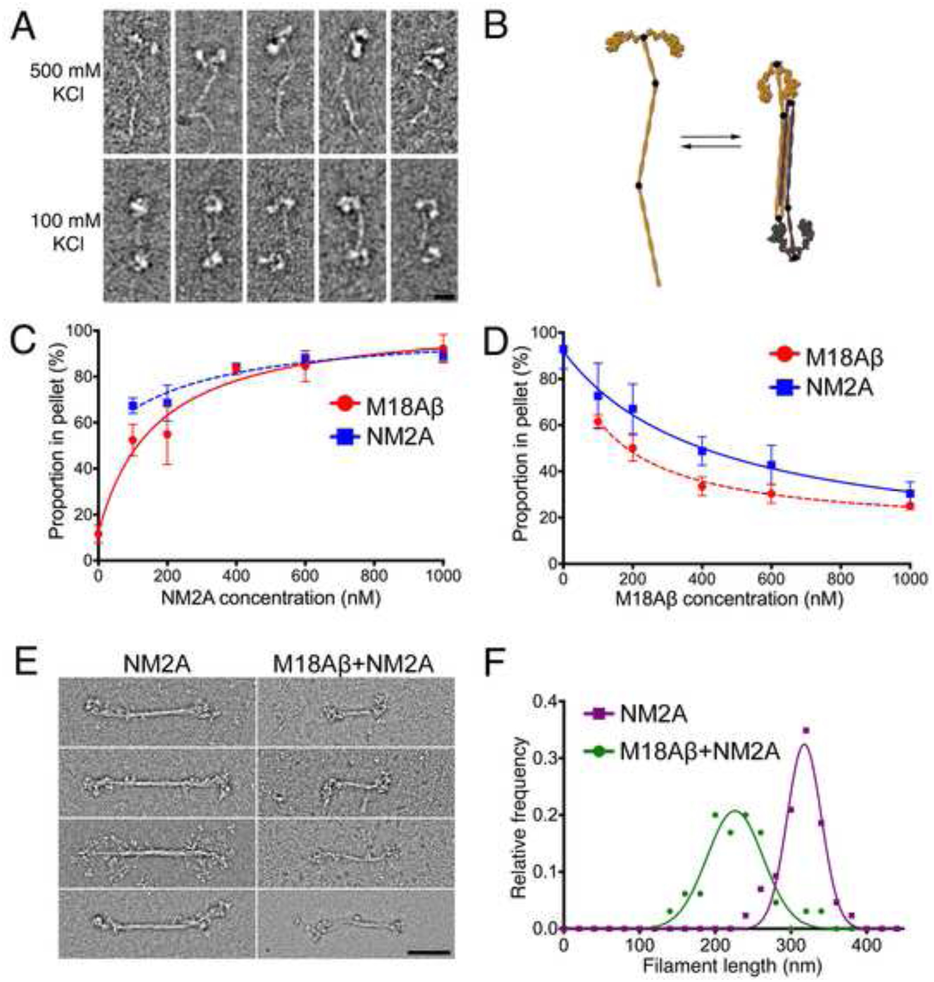
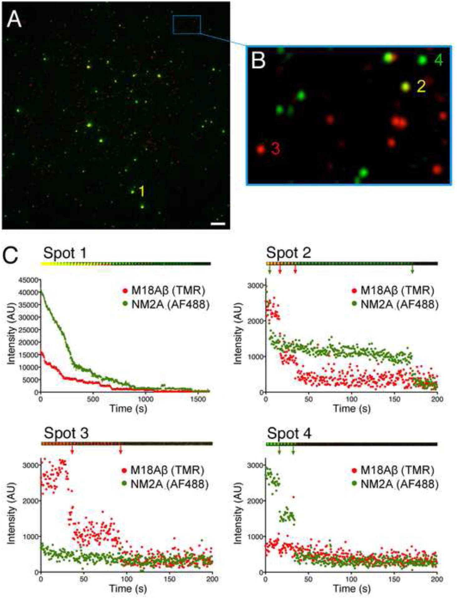
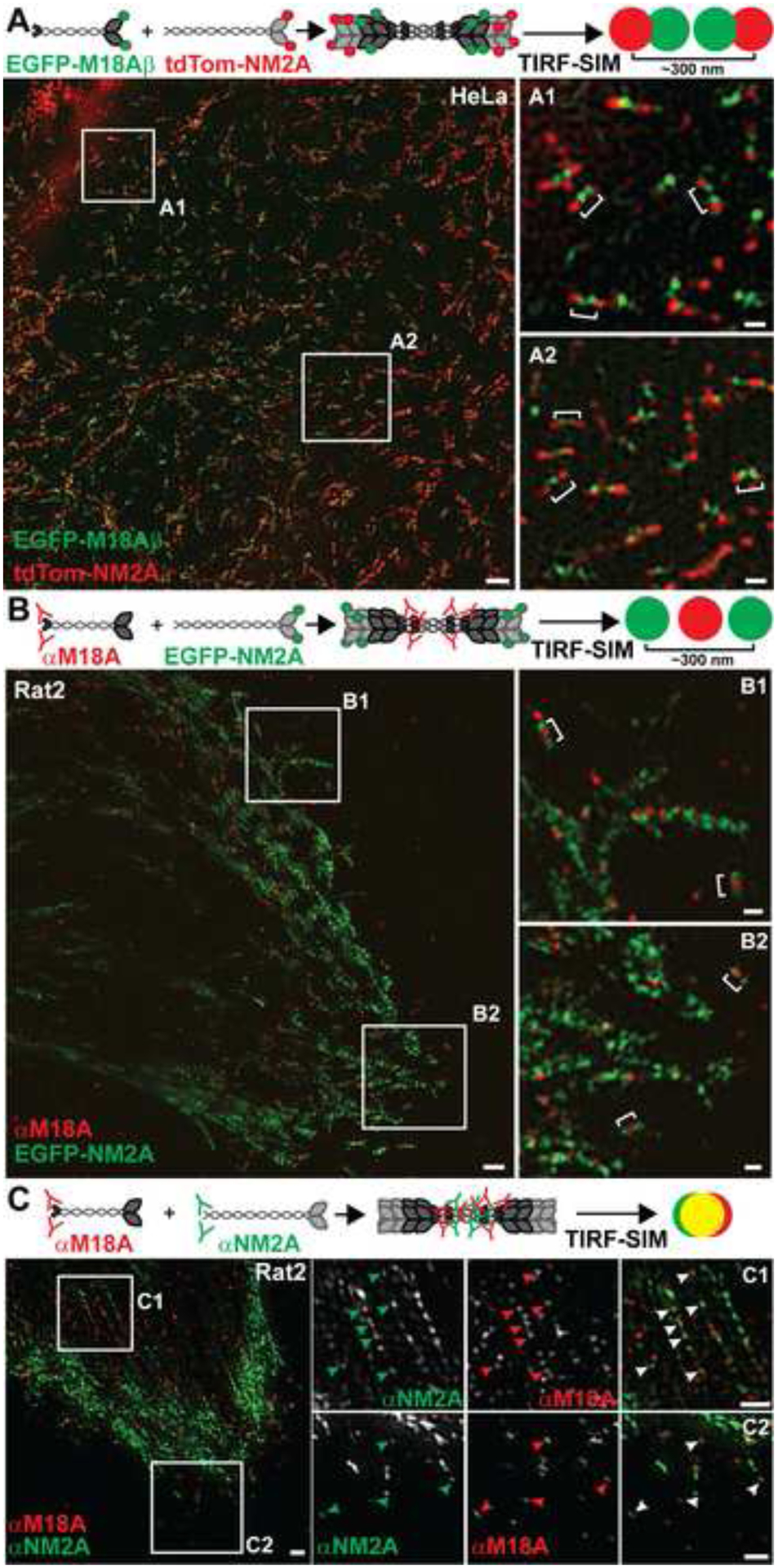
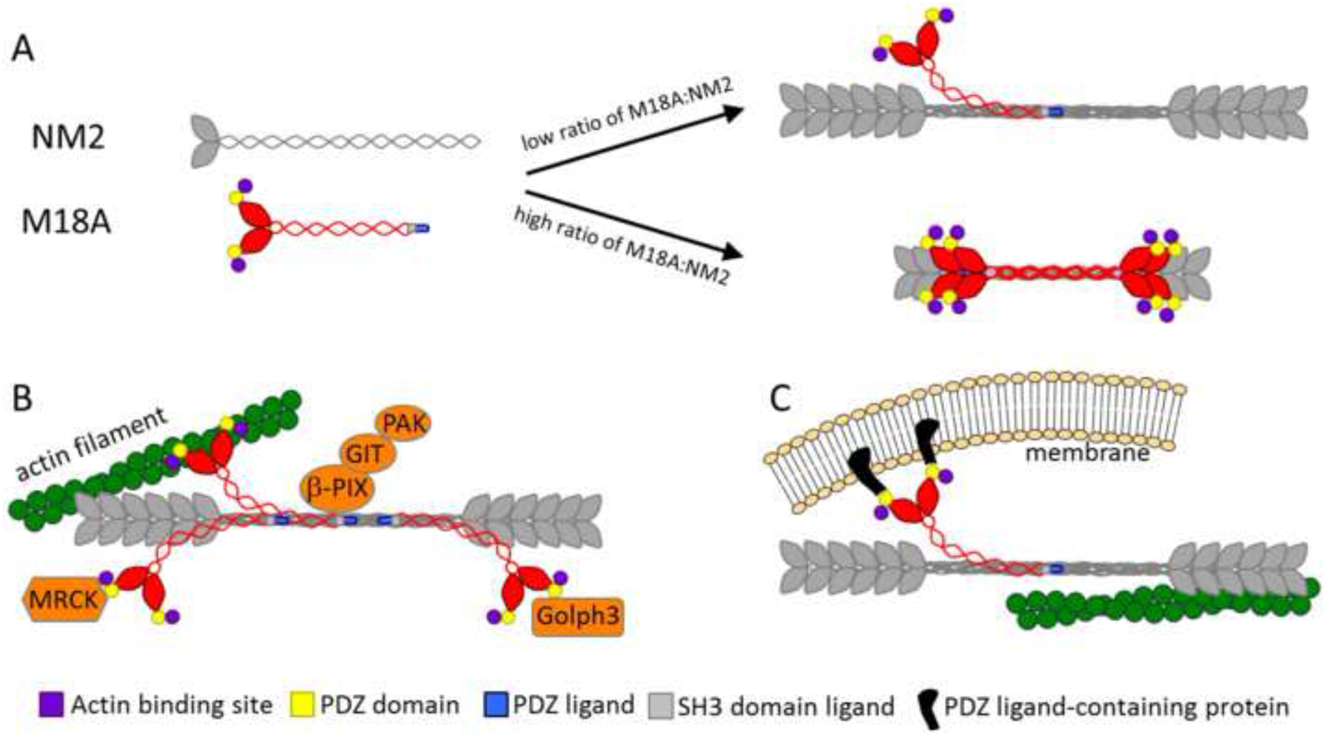
Class-18 myosins are most closely related to conventional class-2 nonmuscle myosins (NM2). Surprisingly, the purified head domains of Drosophila, mouse, and human myosin 18A (M18A) lack actin-activated ATPase activity and the ability to translocate actin filaments, suggesting that the functions of M18A in vivo do not depend on intrinsic motor activity. M18A has the longest coiled coil of any myosin outside of the class-2 myosins, suggesting that it might form bipolar filaments similar to conventional myosins. To address this possibility, we expressed and purified full-length mouse M18A using the baculovirus/Sf9 system. M18A did not form large bipolar filaments under any of the conditions tested. Instead, M18A formed an ∼ 65-nm-long bipolar structure with two heads at each end. Importantly, when NM2 was polymerized in the presence of M18A, the two myosins formed mixed bipolar filaments, as evidenced by cosedimentation, electron microscopy, and single-molecule imaging. Moreover, super-resolution imaging of NM2 and M18A using fluorescently tagged proteins and immunostaining of endogenous proteins showed that NM2 and M18A are present together within individual filaments inside living cells. Together, our in vitro and live-cell imaging data argue strongly that M18A coassembles with NM2 into mixed bipolar filaments. M18A could regulate the biophysical properties of these filaments and, by virtue of its extra N- and C-terminal domains, determine the localization and/or molecular interactions of the filaments. Given the numerous, fundamental cellular and developmental roles attributed to NM2, our results have far-reaching biological implications.


Figures

Figure 1.
Figure 1.. M18A and NM2A form heteropolymers
(A) EM images of M18Aβ in high salt buffer (500 mM KCl) (upper row) and low salt buffer (100 mM KCl) (lower row). Scale bar: 20 nm. (B) Model showing proposed antiparallel interaction between two folded M18Aβ molecules. (C) Cosedimentation of a fixed concentration of M18Aβ (200 nM) with a range of NM2A concentrations; error bars denote S.D. (D) Cosedimentation of a fixed concentration of NM2A (200 nM) with a range of M18Aβ concentrations; error bars denote S.D. (E) EM images comparing filaments of pure NM2A (left column) with filaments from an equimolar mixture of M18Aβ and NM2A (150 mM KCl; right column). Scale bar: 100 nm. (F) Length distribution for NM2A filaments alone (purple; n=43) and a 1:1 mixture of M18Aβ and NM2A (green; n=65), measured from electron micrographs.
Figure 2.
Figure 2.. TIRF microscopy of M18A-NM2A copolymers
(A) TIRF image showing copolymers of M18Aβ:NM2A (e.g. spot 1). (B) Magnified inset showing low intensity puncta consistent with small M18A:NM2A oligomers (e.g. spot 2) and single molecules of M18A (e.g. spot 3) or NM2A (e.g. spot 4). (C) Photobleaching traces of spots highlighted in (A) and (B). Montages above each plot show images of denoted spots over time. Each image of spot 1 shows the average of a 30 second bin, each image of spots 2, 3 and 4 shows the average of a 4 second bin. Arrows denote photobleaching events.
Figure 3.
Figure 3.. M18A and NM2 coassemble in living cells
(A) Top: Cartoon depicting coassembly of EGFP-M18Aβ with tdTom-NM2A and the resulting red-green-green-red pattern observed in TIRF-SIM. Bottom: TIRF-SIM image of HeLa cells expressing EGFP-M18Aβ and tdTom-NM2A. White numbered boxes correspond to the magnified insets. White brackets indicate heterotypic filaments of M18A and NM2A. Occasionally, red puncta are bifurcated by a single green punctum. This is probably due to merging of the two green puncta since the distance between M18A heads is close to the axial resolution of TIRF-SIM. Indeed, some slight offset in green and red puncta is normal at this resolution [16]. Scale bars represent 2 μm for the larger image and 300 nm for insets. (B) Top: Cartoon depicting coassembly of EGFP-NM2A with endogenous M18A localized with an antibody to its C-terminus, and the resulting green-red-green pattern observed in TIRF-SIM. Bottom TIRF-SIM image of Rat2 cells expressing EGFP-NM2A and immunostained for M18A (red secondary antibody). White numbered boxes correspond to the magnified insets. White brackets indicate mixed filaments of M18A and NM2A. Scale bars as in (A). (C) Top: Cartoon depicting coassembly of endogenous M18A with endogenous NM2A both localized with antibodies to their respective C-termini, and the yellow puncta with various amounts of closely-associated red and green signals observed in TIRF-SIM. Bottom: Rat2 cells immunostained for endogenous M18A (red secondary antibody) and NM2A (primary antibody directly conjugated to green Alexa FluorAF488) were imaged with TIRF-SIM. Numbered boxes correspond to the row of images on the right, which are presented as gray scale images in the red and green channels and color in the overlay channel. Arrowheads indicate some of the overlapping green and red puncta indicative of mixed filaments. Note that yellow in the overlay channel is only very obvious when the intensities for red and green are approximately equal, which is often not the case when examining the split images. Scale bars represent 1 μm.
Figure 4.
Figure 4.. Models showing potential roles for M18:NM2 coassembly in vivo
(A) The addition of M18A to NM2 filaments may regulate filament size and mechanochemistry. Based on our in vitro data, the extent to which M18A effects NM2 filament size depends on the relative concentrations of the two myosins, with low ratios of M18A to NM2 (probably the most common situation in vivo) having minimal effects, and with high ratios of M18A to NM2 significantly reducing filament size and likely force output. (B) Addition of M18A, with its protein: protein interaction domains, to NM2 filaments may allow recruitment of specific molecules to hybrid filaments. (C) These protein: protein interaction domains may also serve to attach hybrid filaments to anchored structures against which the filaments can then generate contractile force.

References

    1. Guzik-Lendrum S, Heissler SM, Billington N, Takagi Y, Yang Y, Knight PJ, Homsher E, and Sellers JR (2013). Mammalian myosin-18A, a highly divergent myosin. J Biol Chem 288, 9532–9548. - PMC - PubMed
    1. Taft MH, Behrmann E, Munske-Weidemann LC, Thiel C, Raunser S, and Manstein DJ (2013). Functional characterization of human myosin-18A and its interaction with F-actin and GOLPH3. J Biol Chem 288, 30029–30041. - PMC - PubMed
    1. Guzik-Lendrum S, Nagy A, Takagi Y, Houdusse A, and Sellers JR (2011). Drosophila melanogaster myosin-18 represents a highly divergent motor with actin tethering properties. J Biol Chem 286, 21755–21766. - PMC - PubMed
    1. Billington N, Wang A, Mao J, Adelstein RS, and Sellers JR (2013). Characterization of three full-length human nonmuscle myosin II paralogs. J Biol Chem 288, 33398–33410. - PMC - PubMed
    1. Lowey S, Slayter HS, Weeds AG, and Baker H (1969). Substructure of the myosin molecule. I. Subfragments of myosin by enzymic degradation. J Mol Biol 42, 1–29. - PubMed

Publication types

LinkOut - more resources