. 2015 Mar 16;25(6):702-712.
doi: 10.1016/j.cub.2015.01.016. Epub 2015 Mar 5.

Sleep interacts with aβ to modulate intrinsic neuronal excitability

Affiliations

Sleep interacts with aβ to modulate intrinsic neuronal excitability

Masashi Tabuchi et al. Curr Biol. .

Abstract

Background: Emerging data suggest an important relationship between sleep and Alzheimer's disease (AD), but how poor sleep promotes the development of AD remains unclear.

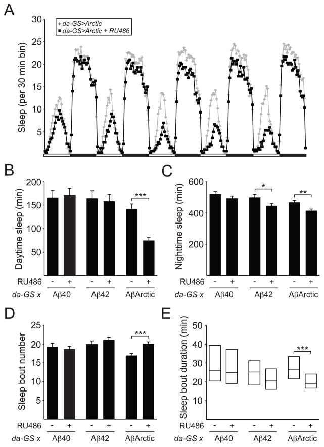
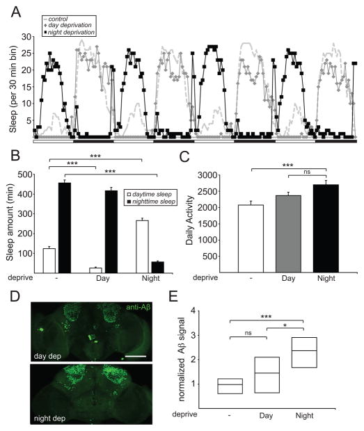
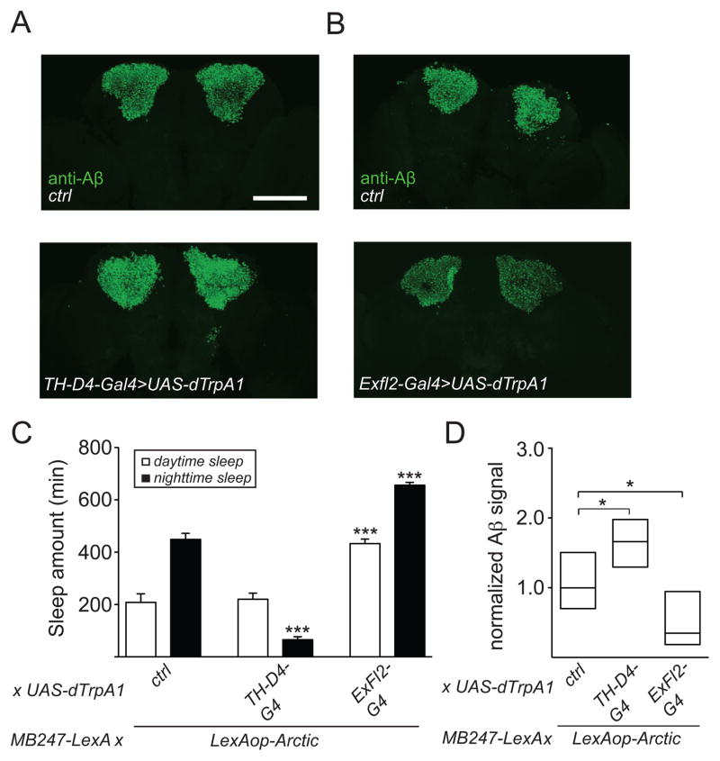
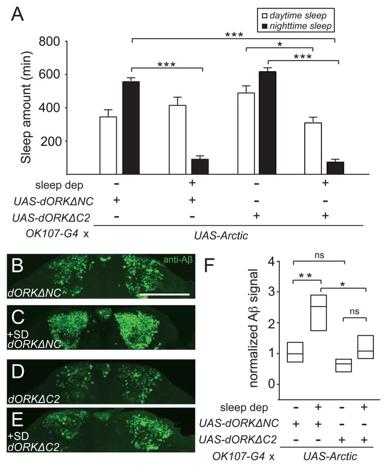
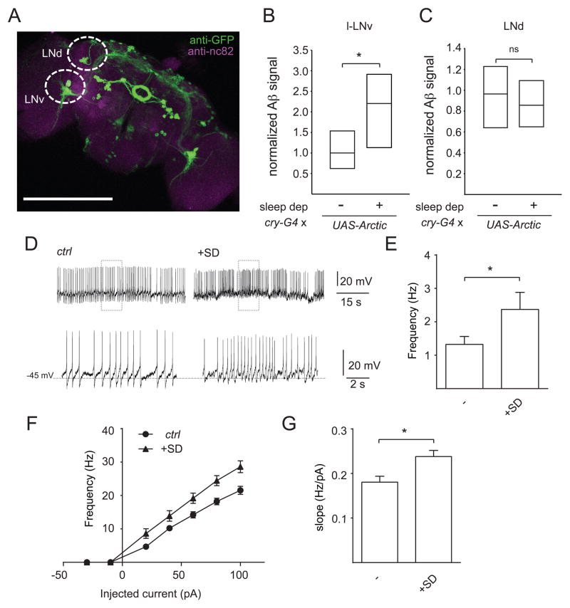
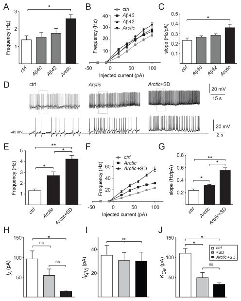
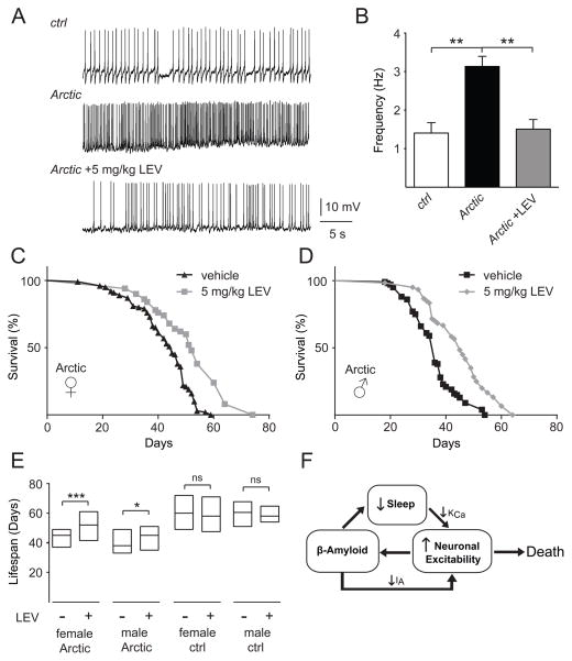
Results: Here, using a Drosophila model of AD, we provide evidence suggesting that changes in neuronal excitability underlie the effects of sleep loss on AD pathogenesis. β-amyloid (Aβ) accumulation leads to reduced and fragmented sleep, while chronic sleep deprivation increases Aβ burden. Moreover, enhancing sleep reduces Aβ deposition. Increasing neuronal excitability phenocopies the effects of reducing sleep on Aβ, and decreasing neuronal activity blocks the elevated Aβ accumulation induced by sleep deprivation. At the single neuron level, we find that chronic sleep deprivation, as well as Aβ expression, enhances intrinsic neuronal excitability. Importantly, these data reveal that sleep loss exacerbates Aβ-induced hyperexcitability and suggest that defects in specific K(+) currents underlie the hyperexcitability caused by sleep loss and Aβ expression. Finally, we show that feeding levetiracetam, an anti-epileptic medication, to Aβ-expressing flies suppresses neuronal excitability and significantly prolongs their lifespan.

Conclusions: Our findings directly link sleep loss to changes in neuronal excitability and Aβ accumulation and further suggest that neuronal hyperexcitability is an important mediator of Aβ toxicity. Taken together, these data provide a mechanistic framework for a positive feedback loop, whereby sleep loss and neuronal excitation accelerate the accumulation of Aβ, a key pathogenic step in the development of AD.


Figures

Figure 1
Figure 1. Induction of AβArctic expression reduces and fragments sleep
(A) Sleep profile for da-GS>UAS-AβArctic flies fed 250 μM RU486 (black squares, n=92) or vehicle control (gray diamonds, n=77). Daytime sleep (B), nighttime sleep (C), nighttime sleep bout number (D), and nighttime sleep bout duration (E) for da-GS>Aβ40 fed RU486 (n=48) or vehicle (n=38), da-GS>Aβ42 fed RU486 (n=70) or vehicle (n=36), and da-GS>AβArctic fed RU486 or vehicle. Data in A are from the same flies as in B–E. In this and subsequent figures, error bars represent SEM. “*”, “**”, “***”, and “ns” denote P<0.05, P<0.01, P<0.001, and not significant, respectively.
Figure 2
Figure 2. Mechanical sleep deprivation enhances Aβ burden
(A) Sleep profile for OK107-Gal4>UAS-AβArctic flies undergoing no, daytime, or nighttime sleep deprivation from a representative experiment. White bars and black bars denote light and dark periods, respectively. Sleep amount (B) and daily activity (C) for OK107-Gal4>UAS-AβArctic flies, where “-”, “Day”, and “Night” denote no, daytime, and nighttime sleep deprivation, respectively. (D) Representative whole-mount brain confocal images for OK107-Gal4>UAS-AβArctic flies undergoing daytime (“day dep”) or nighttime (“night dep”) sleep deprivation, immunostained with anti-Aβ42 antibody (6E10). Maximum projection images are shown. (E) Normalized Aβ signal intensity in the MB KC from OK107-Gal4>UAS-AβArctic flies undergoing no (n=10), daytime (n=10), or nighttime (n=9) sleep deprivation. Aβ signal intensity is not normally distributed and is thus presented here and in subsequent figures as a simplified box plot with the median shown as the line inside the box, and the 75th and 25th percentiles shown as the top and bottom, respectively. Scale bar represents 100 μm.
Figure 3
Figure 3. Genetic manipulation of sleep modulates Aβ levels
(A and B) Representative maximum projection images of whole-mount brains immunostained with 6E10 from LexAop-AβArctic/+; UAS-dTrpA1/MB247-LexA (“ctrl,” top panels) and LexAop-AβArctic/+; UAS-dTrpA1/MB247-LexA, TH-D4-Gal4 (“TH-D4-Gal4>UAS-dTrpA1,” bottom panel), and LexAop-AβArctic/+; UAS-dTrpA1/R72G06-Gal4, MB247-LexA (“ExFl2-Gal4>UAS-dTrpA1,” bottom panel) flies. (C) Daytime and nighttime sleep amounts for LexAop-AβArctic/+; UAS-dTrpA1/MB247-LexA (n=24), LexAop-AβArctic/+; UAS-dTrpA1/MB247-LexA, TH-D4-Gal4 (n=24), and LexAop-AβArctic/+; UAS-dTrpA1/R72G06-Gal4, MB247-LexA (n=22). (D) Normalized Aβ signal intensity in the MB KC for the flies in (C), shown as a simplified box plot. dTrpA1 was chronically activated by subjecting flies to 29°C for 1 week. Scale bar represents 100 μm.
Figure 4
Figure 4. Inhibiting neuronal excitability suppresses Aβ accumulation induced by sleep loss
Daytime and nighttime sleep amounts (A), representative maximum projection images of KC from whole-mount brains immunostained with 6E10 (B–E), and normalized Aβ signal intensity in the MB KC neurons (F) for OK107-Gal4>UAS-AβArctic, UAS-dORKΔNC without sleep deprivation (n=20) and with sleep deprivation (n=13) and OK107-Gal4>UAS-AβArctic, UAS-dORKΔC2 without sleep deprivation (n=17) and with sleep deprivation (n=13). Scale bar represents 100 μm.
Figure 5
Figure 5. Sleep deprivation increases intrinsic neuronal excitability
(A) Maximum projection of a whole-mount brain immunostained with anti-GFP from cry-Gal4>UASCD8:: GFP. Normalized Aβ signal intensity in l-LNv (B) and LNd (C) cells for cry-Gal4>UAS-AβArctic with (n=24) or without sleep deprivation (n=21), shown as a simplified box plot. (D) Representative traces showing spontaneous AP firing of l-LNvs at ZT0-3 in cry-Gal4>UAS-CD8::GFP flies with or without sleep deprivation (SD). The bottom traces in (D) are expanded traces of the boxed regions in the top traces. Mean firing rate of spontaneous activity (E), mean frequency of spikes elicited in response to current injections with 300 ms stepping pulses at 20 pA increments, ranging from −30 pA to 100 pA (F), and f-I slope (G) of l-LNv neurons in control (cry-Gal4>UAS-CD8::GFP) animals with (n=12) or without (n=15) sleep deprivation. Recordings were performed in the presence of mecamylamine (50 μM) and picrotoxin (250 μM), in order to isolate these cells from most excitatory and inhibitory inputs. Scale bar represents 200 μm.
Figure 6
Figure 6. Sleep deprivation exacerbates Aβ-dependent neuronal hyperexcitability
Mean firing rate of spontaneous activity (A), mean frequency of spikes elicited in response to current injections ranging from −30 pA to 100 pA (B), and f-I slope (C) of l-LNv neurons in control cry-Gal4>UAS-CD8::GFP (n=16), cry-Gal4>UAS-Aβ40, UAS-CD8::GFP (n=17), cry-Gal4>UAS-Aβ42, UAS-CD8::GFP (n=15), and cry-Gal4>UAS-AβArctic, UAS-CD8::GFP (n=18). (D) Representative traces showing AP firing of l-LNv neurons in control vs cry-Gal4>UAS-AβArctic, UAS-CD8::GFP flies +/− sleep deprivation (SD). Bottom traces in (D) are expanded traces of the boxed regions in the top traces. Mean firing rate of spontaneous activity (E), mean frequency of spike elicited in response to current injections ranging from −30 pA to 100 pA (F), and f-I slope (G) of l-LNv neurons in control (n=17) vs cry-Gal4>UAS-AβArctic, UAS-CD8::GFP with (n=15) or without sleep deprivation (n=19). IA (H), IK(V) (I), and KCa (J) current amplitude at the spike threshold (−30 mV) from l-LNvs for cry-Gal4>UAS-CD8::GFP with (n=5, 10, and 11, respectively) or without (n=5, 7, and 8, respectively) sleep deprivation and cry-Gal4>UAS-AβArctic, UAS-CD8::GFP with sleep deprivation (n=5, 6, and 4, respectively). Recordings were performed in the presence of mecamylamine (50 μM) and picrotoxin (250 μM), in order to isolate these cells from most excitatory and inhibitory inputs.
Figure 7
Figure 7. Levetiracetam suppresses neuronal firing and prolongs lifespan of Aβ Arctic-expressing flies
(A) Representative traces showing spontaneous firing of l-LNv neurons in control (cry-Gal4>UASCD8:: GFP) vs cry-Gal4>UAS-AβArctic, UAS-CD8::GFP flies fed vehicle or 5 mg/kg levetiracetam (LEV). (B) Quantification of mean firing rates shown in (A) (n = 4 for control, n=5 for AβArctic, and n=8 for AβArctic + LEV). Survivorship curves of elav-Gal4>UAS-AβArctic female (C) and male (D) flies fed vehicle or 5 mg/kg LEV. (E) Lifespan extension of elav-Gal4>UAS-AβArctic female and male flies by LEV, where lifespan is displayed as a simplified box-plot. Data for the elav-Gal4>UAS-AβArctic (“Arctic”) flies shown here are the same as in (C) and (D) (n=98 for vehicle- and n=52 for LEV-fed females, and n=100 for vehicle- and n=60 for LEV-fed males). For elav-Gal4/+ (“ctrl”) flies, n=30 for vehicle- and LEV-fed females and males. (F) Model connecting sleep, neuronal excitability, and Aβ. Sleep loss leads to a reduction in Ca2+-dependent K+ currents, causing neuronal hyperexcitability. This enhanced excitability, in turn, results in increased Aβ accumulation. Aβ itself reduces sleep and further increases neuronal excitability via a decrease in voltage-gated K+ currents, generating a positive feedback loop whereby sleep loss and Aβ interact to substantially increase neuronal activity and Aβ burden. Increased neuronal excitability then contributes to reduced lifespan.

References

    1. Tarawneh R, Holtzman DM. The clinical problem of symptomatic Alzheimer disease and mild cognitive impairment. Cold Spring Harb Perspect Med. 2012;2:a006148. - PMC - PubMed
    1. Selkoe DJ. Alzheimer’s disease. Cold Spring Harb Perspect Biol. 2011;3:a004457. - PMC - PubMed
    1. Ju YE, Lucey BP, Holtzman DM. Sleep and Alzheimer disease pathology--a bidirectional relationship. Nat Rev Neurol. 2014;10:115–119. - PMC - PubMed
    1. Moe KE, Vitiello MV, Larsen LH, Prinz PN. Sleep/wake patterns in Alzheimer’s disease: relationships with cognition and function. J Sleep Res. 1995;4:15–20. - PubMed
    1. Prinz PN, Vitaliano PP, Vitiello MV, Bokan J, Raskind M, Peskind E, Gerber C. Sleep, EEG and mental function changes in senile dementia of the Alzheimer’s type. Neurobiol Aging. 1982;3:361–370. - PubMed

Publication types

MeSH terms