. 2015 Aug;30(4):307-22.
doi: 10.1111/omi.12095. Epub 2015 Apr 20.

Candida-streptococcal mucosal biofilms display distinct structural and virulence characteristics depending on growth conditions and hyphal morphotypes

Affiliations

Candida-streptococcal mucosal biofilms display distinct structural and virulence characteristics depending on growth conditions and hyphal morphotypes

M M Bertolini et al. Mol Oral Microbiol. 2015 Aug.

Abstract

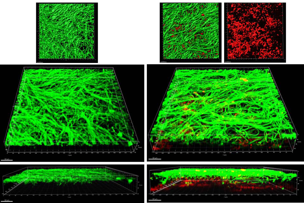
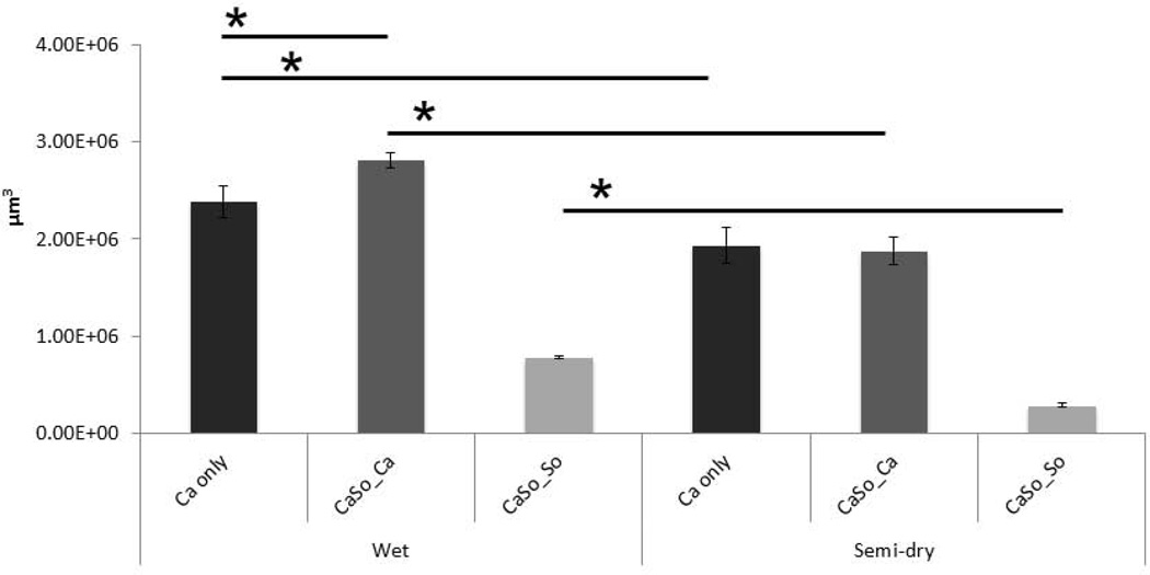
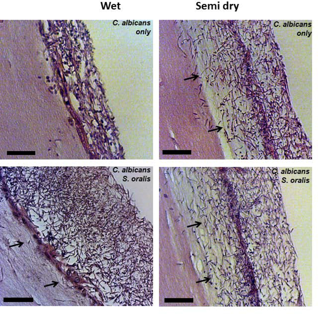
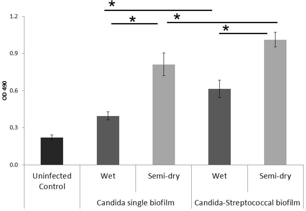
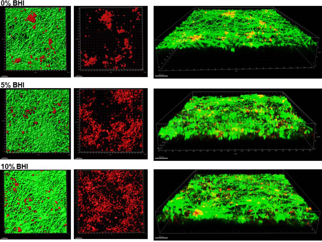
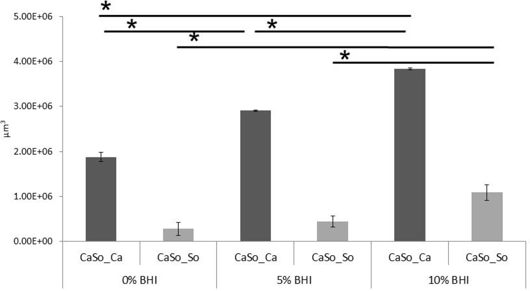
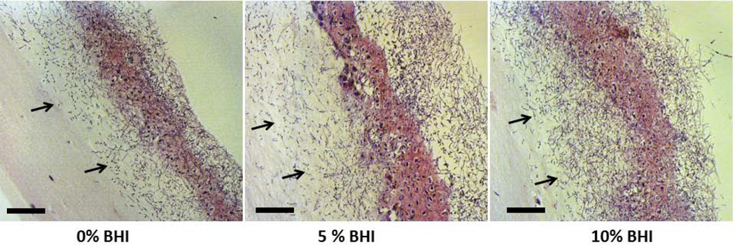
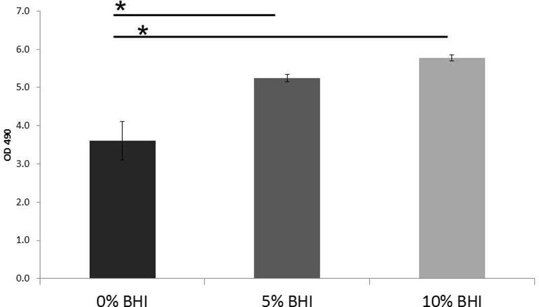
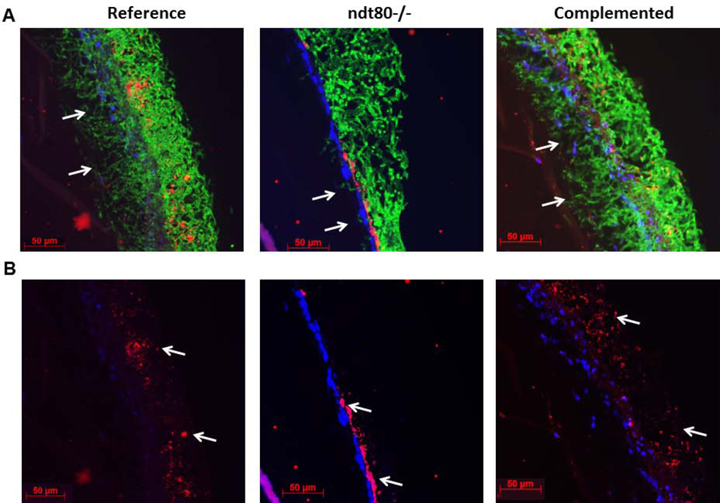
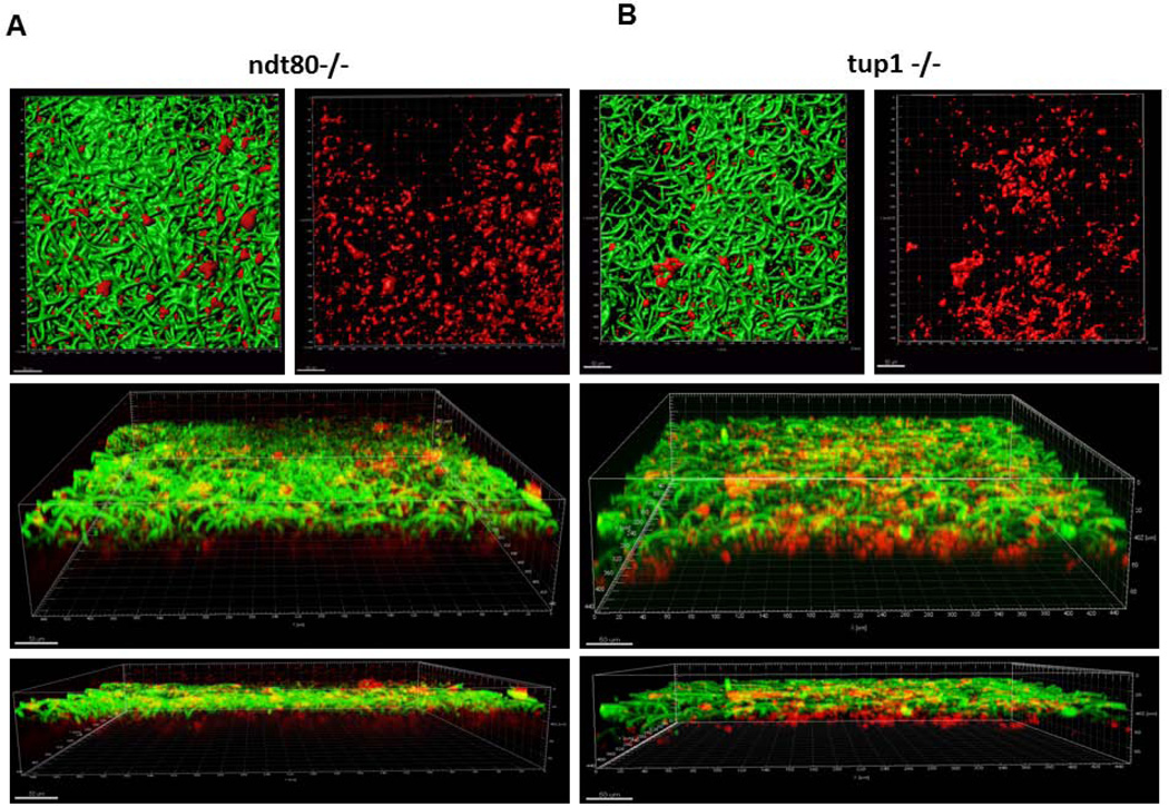
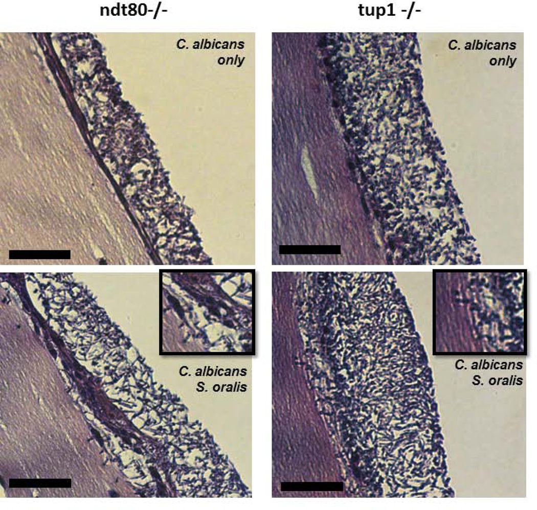
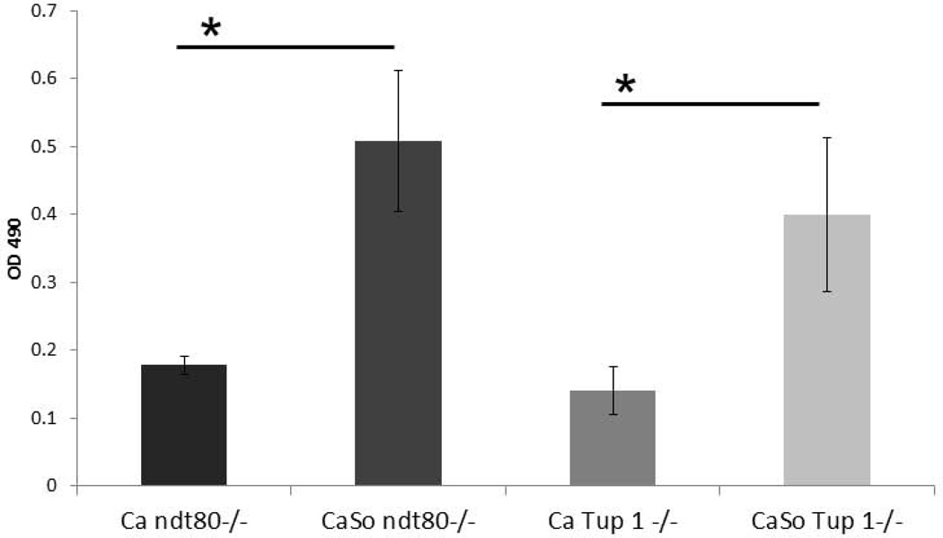
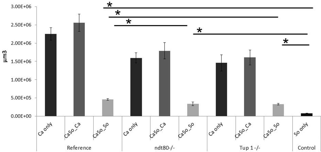
Candida albicans and streptococci of the mitis group form communities in multiple oral sites, where moisture and nutrient availability can change spatially or temporally. This study evaluated structural and virulence characteristics of Candida-streptococcal biofilms formed on moist or semidry mucosal surfaces, and tested the effects of nutrient availability and hyphal morphotype on dual-species biofilms. Three-dimensional models of the oral mucosa formed by immortalized keratinocytes on a fibroblast-embedded collagenous matrix were used. Infections were carried out using Streptococcus oralis strain 34, in combination with a C. albicans wild-type strain, or pseudohyphal-forming mutant strains. Increased moisture promoted a homogeneous surface biofilm by C. albicans. Dual biofilms had a stratified structure, with streptococci growing in close contact with the mucosa and fungi growing on the bacterial surface. Under semidry conditions, Candida formed localized foci of dense growth, which promoted focal growth of streptococci in mixed biofilms. Candida biofilm biovolume was greater under moist conditions, albeit with minimal tissue invasion, compared with semidry conditions. Supplementing the infection medium with nutrients under semidry conditions intensified growth, biofilm biovolume and tissue invasion/damage, without changing biofilm structure. Under these conditions, the pseudohyphal mutants and S. oralis formed defective superficial biofilms, with most bacteria in contact with the epithelial surface, below a pseudohyphal mass, resembling biofilms growing in a moist environment. The presence of S. oralis promoted fungal invasion and tissue damage under all conditions. We conclude that moisture, nutrient availability, hyphal morphotype and the presence of commensal bacteria influence the architecture and virulence characteristics of mucosal fungal biofilms.

Keywords: Candida; Streptococcus; biofilms; infection models.


Figures

Figure 1
Figure 1
A: Sixteen-hour mucosal biofilms of C. albicans monospecies (left panel) or C. albicans-streptococci mixed-species (right panel). Biofilms were grown on the surface of three-dimensional models of oral mucosa under wet (media-submerged) conditions. X-Y isosurfaces (top panel) and 3-D reconstructions (bottom panels) of representative confocal laser scanning microscopy images are shown. C. albicans (green) was visualized after staining with a FITC-conjugated anti-Candida antibody. S. oralis (red) was visualized after fluorescence in situ hybridization (FISH) with a Streptococcus-specific probe conjugated to Alexa 546. Scale bar = 50 µm. B: Sixteen-hour mucosal biofilms of C. albicans monospecies (left panel) or C. albicans-streptococci mixed-species (right panel). Biofilms were grown on the surface of three-dimensional models of oral mucosa under semidry (media limited to inoculum) conditions. X-Y isosurfaces (top panel) and 3-D reconstructions (bottom panels) of representative confocal laser scanning microscopy images are shown. C. albicans (green) was visualized after staining with a FITC-conjugated anti-Candida antibody. S. oralis (red) was visualized after fluorescence in situ hybridization (FISH) with a Streptococcus-specific probe conjugated to Alexa 546. Scale bar = 50 µm.
Figure 1
Figure 1
A: Sixteen-hour mucosal biofilms of C. albicans monospecies (left panel) or C. albicans-streptococci mixed-species (right panel). Biofilms were grown on the surface of three-dimensional models of oral mucosa under wet (media-submerged) conditions. X-Y isosurfaces (top panel) and 3-D reconstructions (bottom panels) of representative confocal laser scanning microscopy images are shown. C. albicans (green) was visualized after staining with a FITC-conjugated anti-Candida antibody. S. oralis (red) was visualized after fluorescence in situ hybridization (FISH) with a Streptococcus-specific probe conjugated to Alexa 546. Scale bar = 50 µm. B: Sixteen-hour mucosal biofilms of C. albicans monospecies (left panel) or C. albicans-streptococci mixed-species (right panel). Biofilms were grown on the surface of three-dimensional models of oral mucosa under semidry (media limited to inoculum) conditions. X-Y isosurfaces (top panel) and 3-D reconstructions (bottom panels) of representative confocal laser scanning microscopy images are shown. C. albicans (green) was visualized after staining with a FITC-conjugated anti-Candida antibody. S. oralis (red) was visualized after fluorescence in situ hybridization (FISH) with a Streptococcus-specific probe conjugated to Alexa 546. Scale bar = 50 µm.
Figure 2
Figure 2
Biovolumes of sixteen-hour single C. albicans (Ca) or mixed C. albicans-S. oralis (CaSo) mucosal biofilms. Average biovolumes were calculated (in µm3) for each species in eight different confocal laser scanning microscopy images using image stacks from two independent experiments. Average biovolumes of C. albicans in single (Ca only) or mixed (CaSo_Ca) biofilms and S. oralis (CaSo_So) in mixed biofilms, under wet (media-submerged) or semidry (media limited to inoculum) conditions. *P = <0.001 when monospecies biovolumes were compared to mixed-species biovolumes or between wet and semi-dry conditions, using the Bonferroni t-test. The error bars indicate one standard deviation of the mean of eight different images from two independent experiments.
Figure 3
Figure 3
H&E-stained tissue sections of 16 hour C. albicans and C. albicans-streptococci mixed-species mucosal biofilms under wet (media-submerged) or semidry (media limited to inoculum) conditions. Arrows indicate invasion of C. albicans through the epithelial barrier formed by OKF6 cells. Scale bar = 50 µm.
Figure 4
Figure 4
Lactate dehydrogenase (LDH) released by mucosal cells. Results represent the average OD490 of subnatant samples from triplicate wells in two independent experiments. *P< 0.05 for a comparison with C. albicans and C. albicans-streptococci mixed-species mucosal biofilms under wet (media-submerged) and semidry (media limited to inoculum) conditions, using the Bonferroni t-test. The error bars indicate one standard deviation of the mean of three different wells from two independent experiments.
Figure 5
Figure 5
A: Sixteen-hour C. albicans-streptococci mixed-species biofilms. Biofilms were grown on the surface of oral mucosa analogues under semidry (media limited to inoculum) conditions and microbial inoculation media were supplemented with 0%, 5% or 10% brain heart infusion broth (BHI). X-Y isosurfaces (left two panels) and 3-D reconstructions (right panel) of representative confocal laser scanning microscopy images of biofilms. C. albicans (green) was visualized after staining with a FITC-conjugated anti-Candida antibody. S. oralis (red) was visualized after fluorescence in situ hybridization (FISH) with a Streptococcus-specific probe conjugated to Alexa 546. Center panel images display the red channel only, showing S. oralis distribution in mixed-species biofilms. Scale bar = 50 µm. B: Average biovolumes (in µm3) for each species in 16h C. albicans. S. oralis (CaSo) mixed-species biofilms grown under the conditions shown in figure 5A. Biovolumes were measured in eight different CLSM image stacks from two independent experiments. Bars represent average biovolumes of C. albicans (CaSo_Ca) or S. oralis (CaSo_So) when grown together. *P = <0.001 when biovolumes were compared between different BHI concentrations, using the Bonferroni t-test. The error bars indicate one standard deviation of the mean of eight different images from two independent experiments.
Figure 5
Figure 5
A: Sixteen-hour C. albicans-streptococci mixed-species biofilms. Biofilms were grown on the surface of oral mucosa analogues under semidry (media limited to inoculum) conditions and microbial inoculation media were supplemented with 0%, 5% or 10% brain heart infusion broth (BHI). X-Y isosurfaces (left two panels) and 3-D reconstructions (right panel) of representative confocal laser scanning microscopy images of biofilms. C. albicans (green) was visualized after staining with a FITC-conjugated anti-Candida antibody. S. oralis (red) was visualized after fluorescence in situ hybridization (FISH) with a Streptococcus-specific probe conjugated to Alexa 546. Center panel images display the red channel only, showing S. oralis distribution in mixed-species biofilms. Scale bar = 50 µm. B: Average biovolumes (in µm3) for each species in 16h C. albicans. S. oralis (CaSo) mixed-species biofilms grown under the conditions shown in figure 5A. Biovolumes were measured in eight different CLSM image stacks from two independent experiments. Bars represent average biovolumes of C. albicans (CaSo_Ca) or S. oralis (CaSo_So) when grown together. *P = <0.001 when biovolumes were compared between different BHI concentrations, using the Bonferroni t-test. The error bars indicate one standard deviation of the mean of eight different images from two independent experiments.
Figure 6
Figure 6
A: H&E-stained tissue sections of 16 h C. albicans-streptococci mixed-species mucosal biofilms. Biofilms were grown on the surface of oral mucosa analogues under semidry (media limited to inoculum) conditions and microbial inoculation media were supplemented with 0%, 5% or 10% BHI. Arrows showing submucosal invasion through the multilayer epithelial barrier formed by SCC15 cells. Scale bar = 50 µm. B: LDH released by oral mucosa analogues with 16 h C. albicans-streptococci mixed-species mucosal biofilms. Biofilms were grown on the surface of oral mucosa analogues under semidry (media limited to inoculum) conditions and microbial inoculation media were supplemented with 0%, 5% or 10% BHI. Results represent the average of OD490 values in triplicate wells from two independent experiments. *P< 0.05 for a comparison with 0% BHI, using the Bonferroni t-test. The error bars indicate one standard deviation of the mean of three different wells from two independent experiments.
Figure 6
Figure 6
A: H&E-stained tissue sections of 16 h C. albicans-streptococci mixed-species mucosal biofilms. Biofilms were grown on the surface of oral mucosa analogues under semidry (media limited to inoculum) conditions and microbial inoculation media were supplemented with 0%, 5% or 10% BHI. Arrows showing submucosal invasion through the multilayer epithelial barrier formed by SCC15 cells. Scale bar = 50 µm. B: LDH released by oral mucosa analogues with 16 h C. albicans-streptococci mixed-species mucosal biofilms. Biofilms were grown on the surface of oral mucosa analogues under semidry (media limited to inoculum) conditions and microbial inoculation media were supplemented with 0%, 5% or 10% BHI. Results represent the average of OD490 values in triplicate wells from two independent experiments. *P< 0.05 for a comparison with 0% BHI, using the Bonferroni t-test. The error bars indicate one standard deviation of the mean of three different wells from two independent experiments.
Figure 7
Figure 7
C. albicans-streptococci mixed-species mucosal biofilms grown under semidry (media limited to inoculum) conditions. C. albicans reference strain, ndt80 homozygous deletion mutant (ndt80−/−) and its complemented strain were grown in the presence of S. oralis 34 for 16h. Representative tissue sections are shown where C. albicans (green) was visualized after staining with a FITC-conjugated anti-Candida antibody and S. oralis (red) was visualized after fluorescence in situ hybridization (FISH) with a Streptococcus-specific probe conjugated to Alexa 546; Mucosal cell nuclei were counterstained with the nucleic acid stain Hoechst 33258 (blue). A: Overlay of three-color images, showing invasion of each Candida strain into the submucosal compartment (white arrows). B: Red and blue channel overlay, showing S. oralis and epithelial nuclei, respectivelly, highlighting the distinct S. oralis biofilm architecture in mutant vs reference and complemented strain biofilms.
Figure 8
Figure 8
C. albicans-streptococci mixed-species mucosal biofilms grown under semidry (media limited to inoculum) conditions. C. albicans pseudohyphal strains, ndt80 homozygous deletion mutant (ndt80−/−) and tup1 homozygous deletion mutant (tup1−/−) were grown in the presence of S. oralis 34 for 16h. Representative x-y isosurfaces (top panel) and 3-D reconstructions (bottom panel) of representative CLSM images. C. albicans (green) was visualized after staining with a FITC-conjugated anti-Candida antibody. S. oralis (red) was visualized after fluorescence in situ hybridization (FISH) with a Streptococcus-specific probe conjugated to Alexa 546. Note the partially stratified structure of biofilms with bacteria co-aggregating on the surface of the oral mucosa as well co-localizing with pseudofilaments (yellow). Scale bar = 50 µm.
Figure 9
Figure 9
A: H&E-stained tissue sections of biofilms shown in figure 8. Magnifying squares show invasion of C. albicans pseudohyphae through the epithelial barrier for both mutant strains. Scale bar = 50 µm. B: LDH released by mucosal analogues inoculated under semidry conditions with pseudohyphal mutants in the presence or absence of S. oralis 34 for 16h. Bars represent the average of the OD490 values in triplicate wells from two independent experiments. *P< 0.05 for a comparison of C. albicans monospecies and C. albicans-streptococci mixed-species mucosal biofilms, using the Bonferroni t-test. The error bars indicate one standard deviation of the mean of three different wells from two independent experiments.
Figure 9
Figure 9
A: H&E-stained tissue sections of biofilms shown in figure 8. Magnifying squares show invasion of C. albicans pseudohyphae through the epithelial barrier for both mutant strains. Scale bar = 50 µm. B: LDH released by mucosal analogues inoculated under semidry conditions with pseudohyphal mutants in the presence or absence of S. oralis 34 for 16h. Bars represent the average of the OD490 values in triplicate wells from two independent experiments. *P< 0.05 for a comparison of C. albicans monospecies and C. albicans-streptococci mixed-species mucosal biofilms, using the Bonferroni t-test. The error bars indicate one standard deviation of the mean of three different wells from two independent experiments.
Figure 10
Figure 10
Average biovolumes (in µm3) for each species in 16h C. albicans-S. oralis (CaSo) mixed-species biofilms grown under the conditions shown in figures 8–9. Biovolumes were measured in eight different CLSM image stacks from two independent experiments. Bars represent average biovolumes of C. albicans (Ca only) single biofilms, S. oralis (So only) single biofilms and C. albicans (CaSo_Ca) or S. oralis (CaSo_So) when grown together. *P = <0.001 when biovolumes were compared between different strains using the Bonferroni t-test. The error bars indicate one standard deviation of the mean of eight different images from two independent experiments.

Similar articles

Cited by

References

    1. Bamford CV, Nobbs AH, Barbour ME, Lamont RJ, Jenkinson HF. Functional regions of Candida albicans hyphal cell wall protein Als3 that determine interaction with the oral bacterium Streptococcus gordonii. Microbiology. 2015;161:18–29. - PMC - PubMed
    1. Brand A, Vacharaksa A, Bendel C, Norton J, Haynes P, Henry-Stanley M, Wells C, Ross K, Gow N, Gale CA. An Internal Polarity Landmark Is Important for Externally Induced Hyphal Behaviors in Candida albicans. Eukaryot Cell. 2008;7:712–720. - PMC - PubMed
    1. Brown DH, Jr., Giusani AD, Chen X, Kumamoto CA. Filamentous growth of Candida albicans in response to physical environmental cues and its regulation by the unique CZF1 gene. Mol. Microbiol. 1999;34:651–662. - PubMed
    1. Cassone A, Cauda R. Candida and candidiasis in HIV-infected patients: where commensalism, opportunistic behavior and frank pathogenicity lose their borders. AIDS. 2012;26:1457–1472. - PubMed
    1. Cavalcanti YW, Morse DJ, da Silva WJ, Del-Bel-Cury AA, Wei X, Wilson M, Milward P, Lewis M, Bradshaw D, Williams DW. Virulence and pathogenicity of Candida albicans is enhanced in biofilms containing oral bacteria. Biofouling. 2015;31:27–38. - PubMed

Publication types