

Percutaneous Distraction Pinning for Metacarpophalangeal Joint Stabilization After Blast or Crush Injuries of the Hand
- PMID: 25754757
- PMCID: PMC4523529
- DOI: 10.1007/s11999-015-4233-x
Percutaneous Distraction Pinning for Metacarpophalangeal Joint Stabilization After Blast or Crush Injuries of the Hand
Abstract
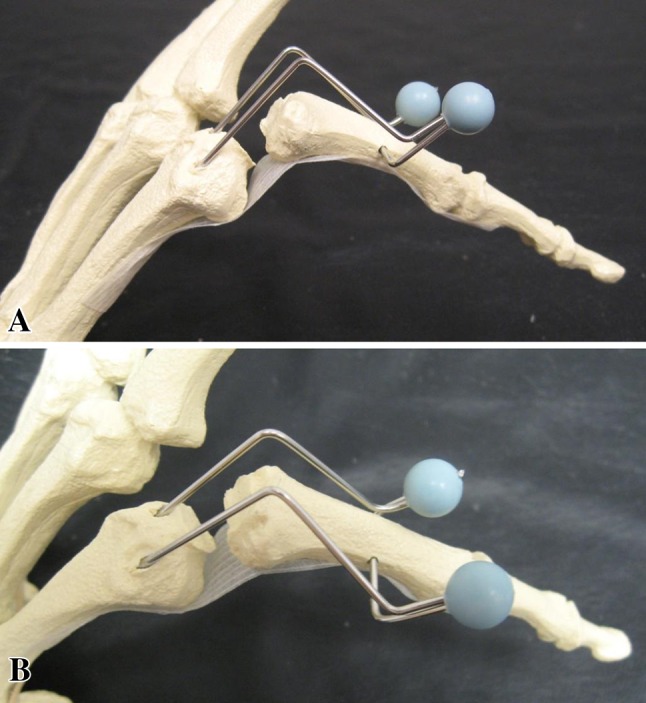
Background: Unstable, severely comminuted fractures of the metacarpophalangeal (MCP) joint are difficult to treat. Closed treatment and casting of these fractures often fail to maintain proper alignment and impede wound care where concomitant open injuries such as gunshot wounds are present. Conventional pinning or plating techniques are not feasible if extensive bone loss and comminution are present. A distraction pinning technique represents a potential alternative, but results with this approach, to our knowledge, have not been reported.
Questions/purposes: The purposes of this study were (1) to evaluate the effectiveness (defined as osseous union and joint stability) of distraction pinning for comminuted fractures involving MCP joints after gunshot or crush injuries; (2) to report the short-term results in terms of pain and function in a small group of patients who underwent MCP distraction pinning; and (3) to evaluate complications and return to work status of these patients in the short term.
Methods: We reviewed 10 patients with comminuted pilon-type fractures of the base of the proximal phalanx or metacarpal head treated with wire distraction fixation from 2005 and 2014. During that period, we used this technique to treat all patients whose fractures were deemed too comminuted for plating or pinning, and during that period, no other techniques (such as simple external fixation) were used for patients meeting those indications. The minimum followup was 6 months; eight of the 10 patients were accounted at a median of 10 months (range, 6-89 months). The median age was 47 years (range, 28-57 years), and seven of the eight were male. Kirschner wire fixation frames were removed 3.5 to 6 weeks after the index surgery when fracture consolidation was confirmed on radiography by the treating surgeon. Stability and range of motion of the MCP joint were assessed using physical examination, radiographs, and goniometer by the treating surgeon. Patients completed the Quick Disabilities of the Arm, Shoulder and Hand score at latest followup or by telephone, and complications were assessed by chart review.
Results: All fractures were healed with stable MCP joints. Eight patients reported having no pain or minimal pain of their injuries to the hand. The median finger and thumb MCP arc of motion were 80° (range, 70°-105°) and 30° (range, 0°-60°), respectively. The median Quick Disabilities of the Arm, Shoulder and Hand score was 3 (range, 0-41). One patient underwent a second surgical procedure for bone grafting and soft tissue coverage. Three patients developed pin site irritations and were treated with oral antibiotics. Six patients returned to their original job.
Conclusions: The distraction pinning technique provides reliable osseous union and joint stability of comminuted pilon-type fractures of the base of the proximal phalanx or metacarpal head, even with associated open wounds. Future studies will need to evaluate these patients at longer term followup and compare this approach with other available techniques, because arthrosis, stiffness, and progressive loss of function seem likely to occur given the severity of these injuries.
Level of evidence: Level IV, therapeutic study.
Figures
References
MeSH terms
LinkOut - more resources
Full Text Sources
Other Literature Sources
Medical
Research Materials
Miscellaneous
