

Outcomes of opening wedge osteotomy to correct angular deformity in little finger clinodactyly
- PMID: 25754787
- PMCID: PMC4410057
- DOI: 10.1016/j.jhsa.2015.01.017
Outcomes of opening wedge osteotomy to correct angular deformity in little finger clinodactyly
Abstract
Purpose: To evaluate the outcomes and complications in a series of children with clinodactyly treated with opening wedge osteotomy of the abnormal phalanx.
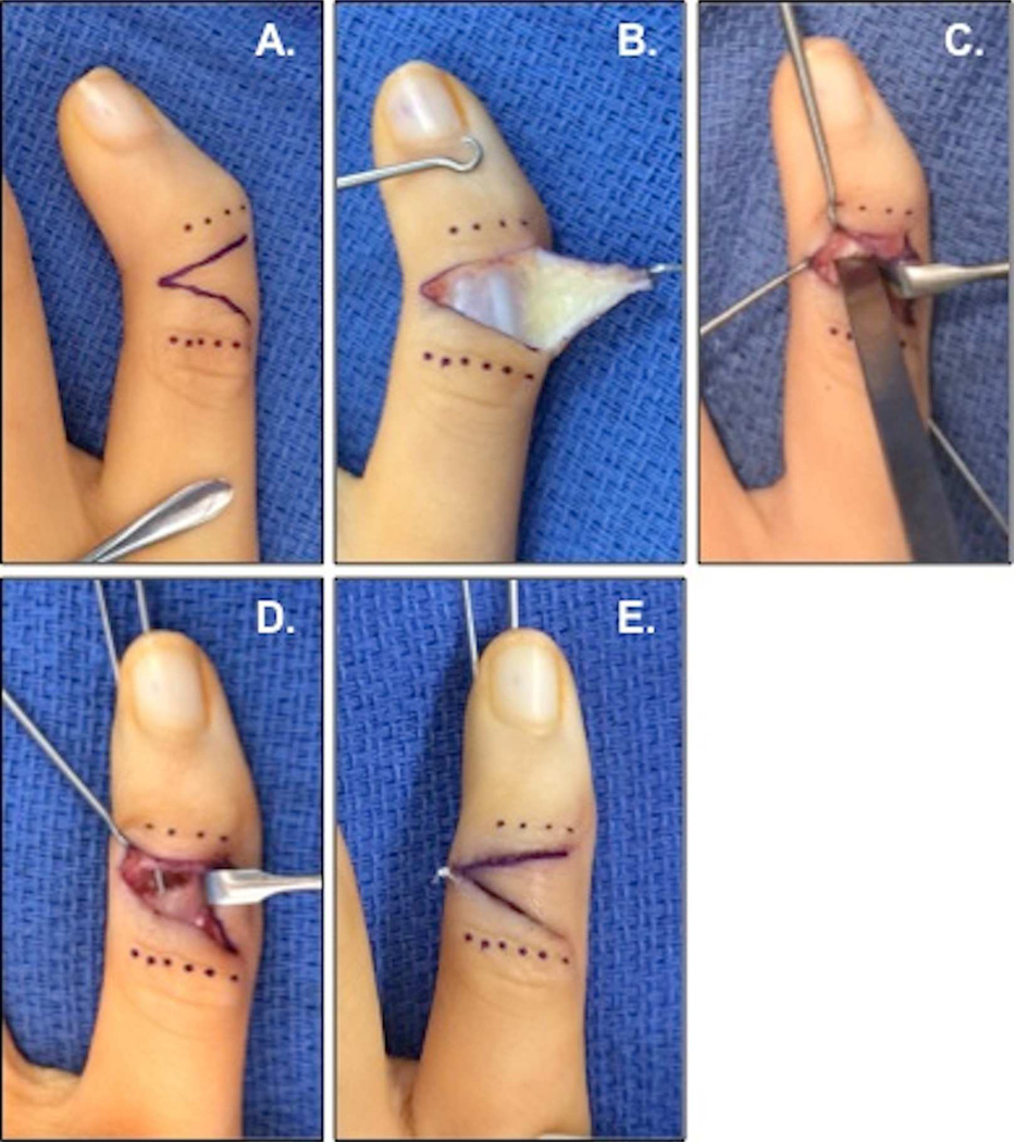
Methods: We performed a retrospective review of all children with clinodactyly treated at our institution with opening wedge osteotomy of the abnormal middle phalanx between 2003 and 2013. Patients with concomitant pathology or prior surgery in the affected finger were excluded. Preoperative and postoperative clinical angle, radiographic angle, digital range of motion, and pain were compared and complications were recorded.
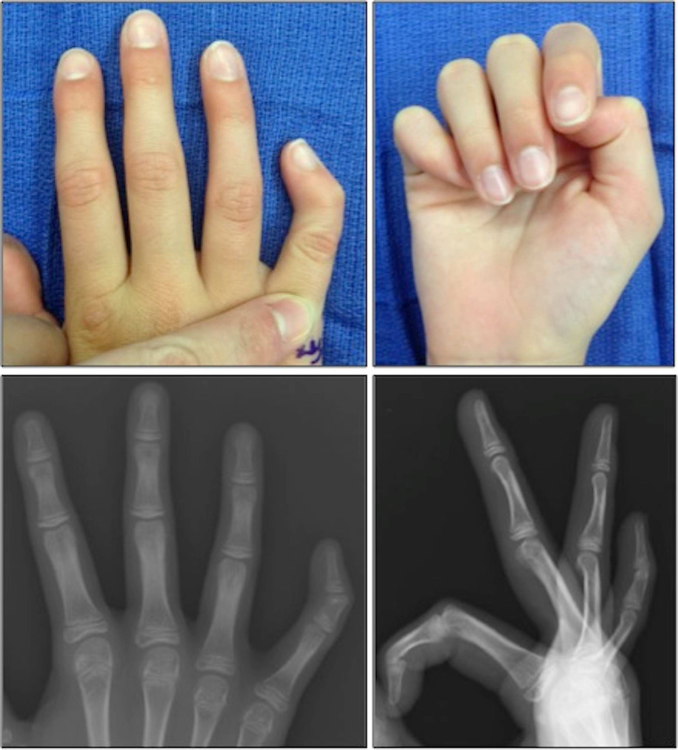
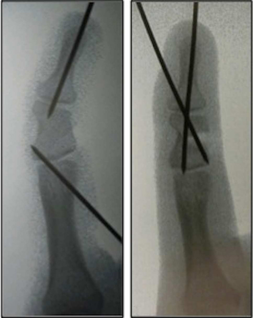
Results: We included 13 digits in 9 patients. All had greater than 20° of preoperative clinical angulation (mean, 36°). Mean age at time of surgery was 11 years; mean duration of follow-up was 25 months (range, 12-43 mo). All digits had significant improvement (mean, 32°) in clinical and radiographic angles after surgery. This improvement was maintained at final follow-up in 12 digits. Six patients had pain preoperatively and no patient had pain postoperatively. One digit had a recurrent deformity at final follow-up and 3 digits developed stiffness at the distal interphalangeal joint.
Conclusions: Opening wedge osteotomy is an effective treatment for angulation in children with clinodactyly. We counsel families regarding the risk of distal interphalangeal joint stiffness.
Type of study/level of evidence: Therapeutic IV.
Keywords: Clinodactyly; opening wedge; osteotomy; surgery.
Copyright © 2015 American Society for Surgery of the Hand. Published by Elsevier Inc. All rights reserved.
Figures
Similar articles
-
Closing wedge osteotomy of abnormal middle phalanx for clinodactyly.J Hand Surg Am. 2009 May-Jun;34(5):914-8. doi: 10.1016/j.jhsa.2009.01.007. Epub 2009 Apr 11. J Hand Surg Am. 2009. PMID: 19362790
-
Long-term results following osteotomy of the thumb delta phalanx in Rubinstein-Taybi Syndrome.J Hand Surg Eur Vol. 2010 May;35(4):296-301. doi: 10.1177/1753193409354523. Epub 2009 Dec 23. J Hand Surg Eur Vol. 2010. PMID: 20031997
-
Lateral Opening-wedge Distal Femoral Osteotomy: Pain Relief, Functional Improvement, and Survivorship at 5 Years.Clin Orthop Relat Res. 2015 Jun;473(6):2009-15. doi: 10.1007/s11999-014-4106-8. Epub 2014 Dec 24. Clin Orthop Relat Res. 2015. PMID: 25537806 Free PMC article.
-
Osteotomy for clinodactyly.J Hand Surg Am. 2015 Jun;40(6):1220-4. doi: 10.1016/j.jhsa.2015.03.003. Epub 2015 Apr 16. J Hand Surg Am. 2015. PMID: 25892713 Review.
-
Malunions of the finger metacarpals and phalanges.Hand Clin. 2006 Aug;22(3):341-55. doi: 10.1016/j.hcl.2006.03.001. Hand Clin. 2006. PMID: 16843800 Review.
Cited by
-
Opening Wedge Osteotomy for Valgus Deformity of the Little Finger after Proximal Phalangeal Fracture in Children: Two Case Reports.Case Rep Orthop. 2018 Apr 17;2018:1526054. doi: 10.1155/2018/1526054. eCollection 2018. Case Rep Orthop. 2018. PMID: 29850327 Free PMC article.
-
Phalangeal Intra-Articular Osteochondroma Caused a Rare Clinodactyly Deformity in Children: Case Series and Literature Review.Front Endocrinol (Lausanne). 2021 Aug 12;12:677245. doi: 10.3389/fendo.2021.677245. eCollection 2021. Front Endocrinol (Lausanne). 2021. PMID: 34456858 Free PMC article. Review.
-
CAMPTODACTYLY AND CLINODACTYLY - NEW UNDERSTANDING OF KNOWN DEFORMITIES.Acta Clin Croat. 2022 Feb;60(3):525-531. doi: 10.20471/acc.2021.60.03.24. Acta Clin Croat. 2022. PMID: 35282479 Free PMC article. Review.
-
A Complex Case of Clino-Syndactyly with Fourth Metacarpal Aplasia.Life (Basel). 2023 Sep 21;13(9):1943. doi: 10.3390/life13091943. Life (Basel). 2023. PMID: 37763346 Free PMC article.
-
Bilobed Flap in Hand Clinodactyly Reconstruction: Technique Description and Result Appraisal.Rev Bras Ortop (Sao Paulo). 2021 Oct 1;57(4):642-648. doi: 10.1055/s-0041-1731797. eCollection 2022 Aug. Rev Bras Ortop (Sao Paulo). 2021. PMID: 35966427 Free PMC article.
References
-
- Light TR, Ogden JA. The longitudinal epiphyseal bracket: implications for surgical correction. J Pediatr Orthop. 1981;1(3):299–305. - PubMed
-
- Ogden JA, Light TR, Conlogue GJ. Correlative roentgenography and morphology of the longitudinal epiphyseal bracket. Skeletal Radiol. 1981;6(2):109–117. - PubMed
-
- Jones GB. Delta Phalanx. J Bone Joint Surg Br. 1964;46:226–228. - PubMed
-
- Burke F, Flatt A. Clinodactyly. A review of a series of cases. Hand. 1979;11(3):269–280. - PubMed
-
- Dutta P. The Inheritance of the Radially Curved Little Finger. Acta Genet Stat Med. 1965;15:70–76. - PubMed
Publication types
MeSH terms
Grants and funding
LinkOut - more resources
Full Text Sources
Other Literature Sources
Research Materials
