. 2015 Mar 18;85(6):1244-56.
doi: 10.1016/j.neuron.2015.02.017. Epub 2015 Mar 5.

Subtype-specific regeneration of retinal ganglion cells following axotomy: effects of osteopontin and mTOR signaling

Affiliations

Subtype-specific regeneration of retinal ganglion cells following axotomy: effects of osteopontin and mTOR signaling

Xin Duan et al. Neuron. .

Abstract

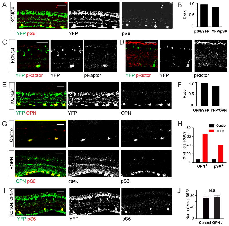
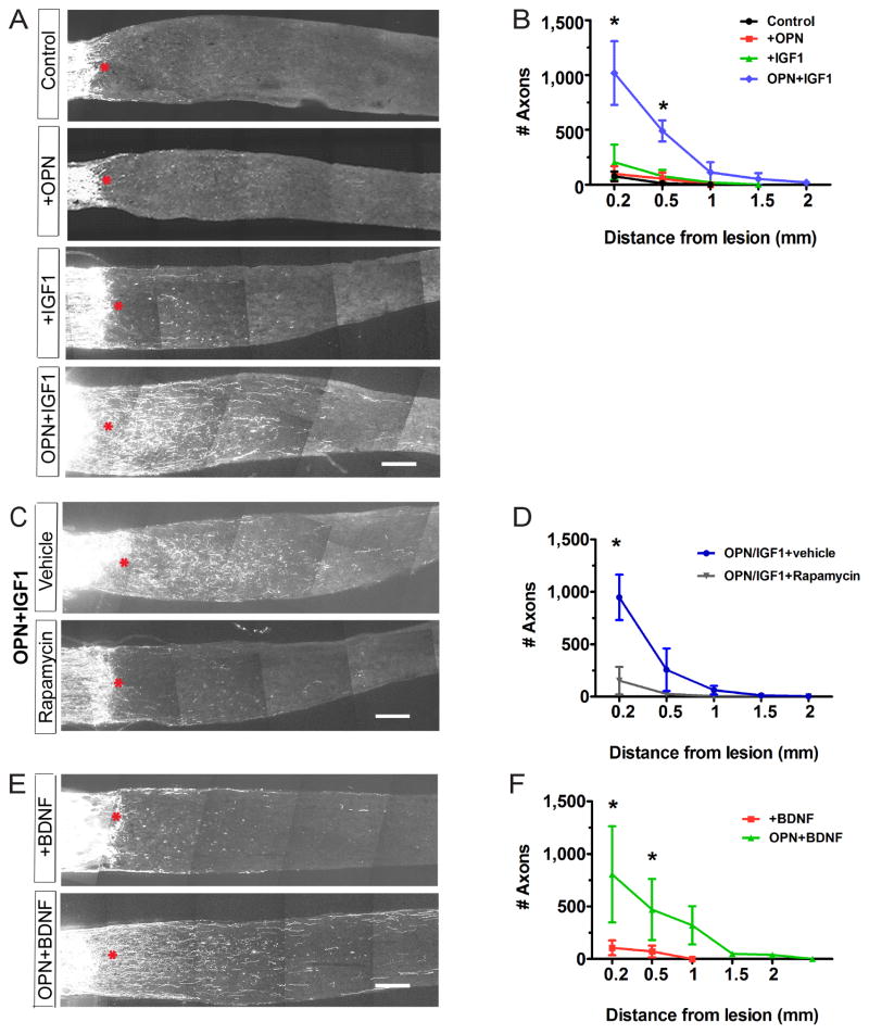
In mammals, few retinal ganglion cells (RGCs) survive following axotomy, and even fewer regenerate axons. This could reflect differential extrinsic influences or the existence of subpopulations that vary in their responses to injury. We tested these alternatives by comparing responses of molecularly distinct subsets of mouse RGCs to axotomy. Survival rates varied dramatically among subtypes, with alpha-RGCs (αRGCs) surviving preferentially. Among survivors, αRGCs accounted for nearly all regeneration following downregulation of PTEN, which activates the mTOR pathway. αRGCs have uniquely high mTOR signaling levels among RGCs and also selectively express osteopontin (OPN) and receptors for the insulin-like growth factor 1 (IGF-1). Administration of OPN plus IGF-1 promotes regeneration as effectively as downregulation of PTEN; however, regeneration is still confined to αRGCs. Our results reveal dramatic subtype-specific differences in the ability of RGCs to survive and regenerate following injury, and they identify promising agents for promoting axonal regeneration.


Figures

Figure 1
Figure 1. Differential survival of RGC subtypes following axotomy
A. Whole-mount views of retinas. Top panels show retina labeled with antibody Tuj1, which marks all RGCs. Lower panels show retinas from Kcng4-YFP, TYW3 and HB9-GFP mice, in which αRGCs, W3-RGCs, and ooDSGCs, respectively, are labeled. dpc, days post-crush. Scale bar, 50μm. B. Retinal sections labeled with anti-melanopsin in wild-type mice to label M1 and M2 cells, and YFP in Kcng4-YFP mice to label αRGCs. 1 and 2 indicate M1 and M2 RGCs, which can be distinguished by dendritic lamination; only M1 RGCs survive. 3–5 indicate examples of ON-, OFF-transient and OFF-sustained αRGCs, which can be distinguished by dendritic lamination; all 3 survive axotomy. Scale bar, 20μm. C, D. Fraction of RGCs of each subtype that survive axotomy at 14dpc (C) or 28dpc (D); data from preparations such as those shown in A and B. n=2–3 retinas per type. E. Fraction of all RGCs comprised by each subtype in intact retina, 14dpc, and 28dpc. (See also Figure S1.)
Figure 2
Figure 2. Selective regeneration of αRGCs following axotomy
A. Sections from control retina, and a retina infected 2 weeks previously with AAV-shPTEN. Sections were stained with anti-pS6 and Tuj1. PTEN knockdown leads to increased mTOR signaling, revealed by increased levels of pS6. Scale bar, 20μm. B. Sections from optic nerves of Kcng4-YFP, TYW3 and HB9-GFP mice at 14dpc and 28 day after injection of AAV-shPTEN. * marks lesion site. Scale bar, 200μm. C. Section from Kcng4-YFP mouse, as in B but injected with cholera toxin B (CTB) to label all regenerated axons. All CTB-positive axons are YFP+ αRGCs. Scale bar, 200μm. D. Number of labeled regenerating axons 0.5mm distal to the lesion site at 14dpc, based on counts from sections such as those shown in (C), mean+/- S.D., n=3–5 optic nerves per type. *, p<0.05 (Two-ANOVA with Bonferroni Posttests). (See also Figure S2.)
Figure 3
Figure 3. Selective mTOR activity and OPN expression in αRGCs
A, B. Section of Kcng4-YFP retina labeled with antibodies to pS6 plus YFP (A) and quantification of their overlap (B). C,D. Sections of Kcng4-YFP retina labeled with antibodies to pRaptor (C) or pRictor (D) plus YFP. E, F. Section of Kcng4-YFP retina labeled with antibodies to OPN and YFP (E) and quantification of their overlap (F). G, H. Section of control retina (top) and retina infected with AAV-OPN (bottom) 2 weeks previously, labeled with antibodies to OPN and pS6 (G). (H) shows fraction of OPN+ and pS6+ cells in both conditions. I, J. Section of Kcng4-YFP;OPN−/− retina labeled with antibodies to pS6 and YFP (I) and fraction of YFP+ cells that were pS6+ (J). n.s., not significant. n=3 retinas per condition. Scale bars are 50μm.
Figure 4
Figure 4. Role of OPN in developing retina
A. Sections from retinas from TYW7 mice of indicated ages stained with antibodies to YFP and OPN. TYW7 labels OFF αRGCs (Kim et al. 2010 and B. Krieger, M.Q, X.D., J.R.S. and M. Meister, in preparation). OPN+ YFP cells are presumably ON αRGCs. Staining is absent in retinas from OPN−/− mice. Scale bar, 50μm. B. Soma sizes of developing αRGCs and non-αRGCs, and OPN levels in αRGCs in developing retina. OPN levels were measured from sections such as those in (A), as described in Methods. C. Retinas from Thy1-cre mice infected 4 weeks previously with Cre-dependent AAV-YFP, with or without Cre-dependent AAV-OPN. Sections were labeled with antibodies to YFP and OPN. Bar, 20μm D. RGC soma area, calculated from images such as those in C. E. Soma size increase of W3-RGCs and αRGCs, measured from sections such as those in Fig. S3B. ** p<0.01. (See also Figure S3.)
Figure 5
Figure 5. OPN promotes regeneration of axotomized RGCs
A. Sections of optic nerves at 14dpc. Retinas were untreated, infected with AAV-OPN, injected with IGF-1 or BDNF, or both infected and injected. B. Numbers of regenerating fibers at indicated distances from lesion site, measured from sections such as those in (A). C. Sections of optic nerves from Kcng4-YFP mice injected with (AAV-OPN+IGF-1) with or without rapamycin. D. Numbers of regenerating fibers, measured from sections such as those in (C) and Fig. S5C. n = 3–5 optic nerves per condition. E. Sections of optic nerves from Kcng4-YFP mice injected with BDNF with or without AAV-OPN. F. Numbers of regenerating fibers measured from sections such as those in (E). Scale Bar for A, C, E, 200μm. *, p<0.05. (See also Figures S4 and S5.)
Figure 6
Figure 6. Osteopontin promotes selective regeneration of αRGCs
A. Sections of optic nerves of Kcng4-YFP, TYW3, and HB9-GFP mice at 14dpc treated with AAV-OPN and IGF-1 and injected with CTB at 12dpc. Right panels show region boxed in top, left panel. B. Numbers of regenerating fibers 0.5 mm from lesion site from sections such as those in F. n = 3–4 optic nerves per type. *, p<0.05. C. Section of optic nerves from Kcng4-YFP mice injected with Cre-dependent AAV-OPN plus IGF-1. D. Numbers of regenerating fibers 0.5 mm from lesion sites, measured from sections such as those in (C). P<0.05 (Two-ANOVA with Bonferroni Posttests). (See also Figure S6.)
Fig. 7
Fig. 7. Selective expression of IGF1R and activation of mTOR signaling in axotomized αRGCs
A. IGF1R expression in αRGCs (labeled with OPN) in control retina, 3dpc and 7dpc. B–D. Sections from mice injected with control vector, AAV-shPTEN or AAV-OPN plus IGF-1. Optic nerves were crushed 14 days later and retinas analyzed 14dpc with anti-pS6 (B), anti-pRaptor (C) or anti-pRictor (D). Scale bars are 50μm. E. Fraction of αRGC and other RGCs (YFP+ and YFP, respectively in Kcng4-YFP) that are pS6+, from sections such as those in (B). n=4-6 retinas per treatment. F. Model showing pathways by which PTEN knock-down or exogenous OPN expression could promote regeneration. a,b,c indicate steps at which differences between αRGC and non-αRGCs could affect their regenerative abilities. Results in A–E implicate steps “a” and “b” as critical differences (see Discussion). (See also Figure S7.)

References

    1. Aguayo AJ, Rasminsky M, Bray GM, Carbonetto S, McKerracher L, Villegas-Perez MP, Vidal-Sanz M, Carter DA. Degenerative and regenerative responses of injured neurons in the central nervous system of adult mammals. Philosophical transactions of the Royal Society of London Series B, Biological sciences. 1991;331:337–343. - PubMed
    1. Ahmed M, Kundu GC. Osteopontin selectively regulates p70S6K/mTOR phosphorylation leading to NF-kappaB dependent AP-1-mediated ICAM-1 expression in breast cancer cells. Molecular cancer. 2010;9:101. - PMC - PubMed
    1. Bellahcene A, Castronovo V, Ogbureke KU, Fisher LW, Fedarko NS. Small integrin-binding ligand N-linked glycoproteins (SIBLINGs): multifunctional proteins in cancer. Nature reviews Cancer. 2008;8:212–226. - PMC - PubMed
    1. Benowitz LI, Popovich PG. Inflammation and axon regeneration. Current opinion in neurology. 2011;24:577–583. - PubMed
    1. Berson DM. Retinal ganglion-cell types and their central projections. In: Allan AK, Basbaum I, Shepard Gordon M, Westheimer Gerald, editors. The Senses: A Comprehensive Reference. San Diego: Academic Press; 2008. pp. 491–520.

Publication types

MeSH terms