. 2015 May;282(10):2045-59.
doi: 10.1111/febs.13259. Epub 2015 Mar 27.

Protein disulfide isomerase as a novel target for cyclopentenone prostaglandins: implications for hypoxic ischemic injury

Affiliations

Protein disulfide isomerase as a novel target for cyclopentenone prostaglandins: implications for hypoxic ischemic injury

Hao Liu et al. FEBS J. 2015 May.

Abstract

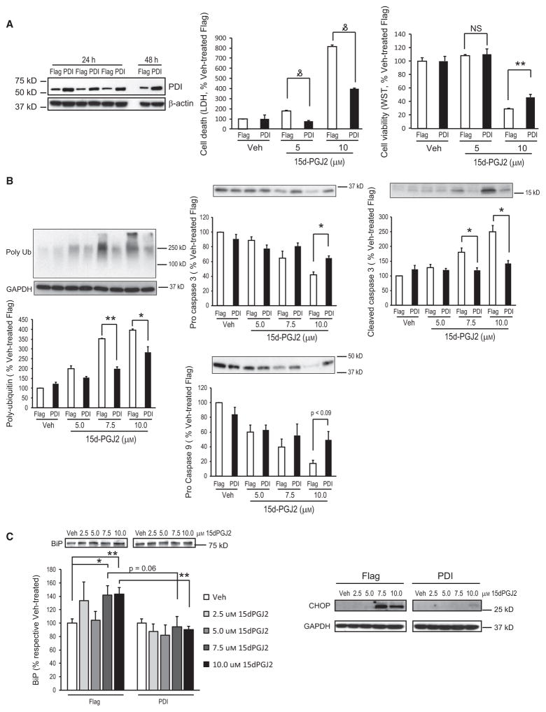
Cyclooxygenase-2 (COX-2) is an important contributor to ischemic brain injury. Identification of the downstream mediators of COX-2 toxicity may allow the development of targeted therapies. Of particular interest is the cyclopentenone family of prostaglandin metabolites. Cyclopentenone prostaglandins (CyPGs) are highly reactive molecules that form covalent bonds with cellular thiols. Protein disulfide isomerase (PDI) is an important molecule for the restoration of denatured proteins following ischemia. Because PDI has several thiols, including thiols within the active thioredoxin-like domain, we hypothesized that PDI is a target of CyPGs and that CyPG binding of PDI is detrimental. CyPG-PDI binding was detected in vitro via immunoprecipitation and MS. CyPG-PDI binding decreased PDI enzymatic activity in recombinant PDI treated with CyPG, and PDI immunoprecipitated from neuronal culture treated with CyPG or anoxia. Toxic effects of binding were demonstrated in experiments showing that: (a) pharmacologic inhibition of PDI increased cell death in anoxic neurons, (b) PDI overexpression protected neurons exposed to anoxia and SH-SY5Y cells exposed to CyPG, and (c) PDI overexpression in SH-SY5Y cells attenuated ubiquitination of proteins and decreased activation of pro-apoptotic caspases. In conclusion, CyPG production and subsequent binding of PDI is a novel and potentially important mechanism of ischemic brain injury. We show that CyPGs bind to PDI, cyclopentenones inhibit PDI activity, and CyPG-PDI binding is associated with increased neuronal susceptibility to anoxia. Additional studies are necessary to determine the relative role of CyPG-dependent inhibition of PDI activity in ischemia and other neurodegenerative disorders.

Keywords: brain; cyclooxygenase; cyclopentenone; ischemia; protein disulfide isomerase.


Figures

Fig. 1
Fig. 1
15d-PGJ2 binds to PDI in vitro and in primary neurons. (A) PDI recombinant protein was preincubated with or without 500 μM 15d-PGJ2 for 30 min before being incubated with vehicle or 5 μM b-15d-PGJ2 for another 90 min. The b-15d-PGJ2–PDI adducts were detected by immunoblotting with streptavidin–HRP. (B) 15d-PGJ2 binds to PDI in primary neurons. (Upper) Rat primary neurons were incubated with 10 μM 15d-PGJ2 or b-15d-PGJ2 for 2 h prior to harvest. The avidin pull-down assay was performed with cell lysates, and b-15d-PGJ2–PDI adducts were detected with PDI antibody. (Lower) Rat primary neurons were incubated with 10 μM b-15d-PGJ2 (+) or vehicle (−) for 2 h before harvest. Cell lysates were either subjected to immunoblotting to detect biotinylated proteins with streptavidin–HRP (Str-H, upper left) and PDI levels with an PDI antibody (lower left) or subjected to IP to detect the b-15d-PGJ2–PDI adduct (right). For IP, cell lysates were incubated with PDI antibody-conjugated resin (R) overnight before elution. A nonreactive control resin was included as a negative control. The b-15d-PGJ2–PDI adduct and PDI in the eluent were detected by immunoblotting with streptavidin–HRP (upper right) and PDI antibody (lower right), respectively. The arrow indicates the band representing b-15d-PGJ2–PDI adduct. (C) Avidin pull-down assay detecting arachidonic acid (AA) metabolite-modified PDI in primary neurons. Neurons were incubated with b-arachidonic acid (b-AA) then underwent hypoxia (+) or normoxia (−) before being harvested at the indicated time points. (Upper) Avidin-bead-bound PDI was detected by immunoblotting with PDI antibody. (Lower) Biotinylated proteins and endogenous PDI in cell lysates were detected by immunoblot with streptavidin–HRP and PDI antibody. (D) Fragmentation spectra of 15d-PGJ2-modified PDI (Uniprot Accession: P07237). The tryptic peptides indicate cysteine (C*) modifications at C-57 or C-60 (upper) and C-401 or C-404 (lower). Cysteine carbamidomethylation from sample processing for mass spectrometry is denoted by C#. MS/MS has an abundance of y- and b-ions N terminal to proline (underlined) with mass shifts corresponding to 316.204 Da indicating 15d-PGJ2 adduction.
Fig. 2
Fig. 2
PDI thiol reductase activity is inhibited by 15d-PGJ2 modification. (A) 15d-PGJ2 reduces recombinant PDI protein thiol reductase activity. Recombinant PDI (5 μM) was incubated with 1.65–33.3 μM of 15d-PGJ2 for 3 h followed by Di-E-GSSG substrate. Thiol reduction was measured by fluorescence plate reader and normalized to untreated recombinant PDI control. Data are means ± SD. n = 2–3 per group. (B) PDI activity is decreased in rat primary neuronal culture after treatment with 15d-PGJ2. Rat primary neuronal cultures were treated with 10 μM vehicle (Veh, methyl acetate) or 10 μM 15d-PGJ2 for 24 h prior to harvest. Neuronal PDI proteins were precipitated with anti-PDI antibody and a thiol reductase activity assay was performed. n = 3 per group. Data are means ± SD. **P < 0.001.
Fig. 3
Fig. 3
Overexpression of PDI decreases 15d-PGJ2-induced cell death in SH-SY5Y cells. SH-SY5Y cells were transfected with Flag–PDI/pcDNA3.1 (PDI, black bar) or control empty vector (Flag, white bar) for 24 h before treatment with 2.5–10 μM 15d-PGJ2 or vehicle (Veh) for an additional 16 h (C) or 24 h (A,B). (A) (Left) Overexpression of PDI in transfected SH-SY5Y cells. SH-SY5Y cells were transfected with Flag–PDI/pcDNA3.1 (PDI) or control empty vector (Flag) then harvested at 24 h (n = 3) and 48 h. Cell lysates were subjected to immunoblotting using anti-PDI antibody, and β-actin was used as loading control. (Center and right) Cell death was measured by LDH assay and cell viability by WST-1 assay. n = 6–12 per group. (B) Representative immunoblots using caspase 9, caspase 3, and poly-ubiquitinated protein antibodies. Densitometric analysis is shown below representative immunoblots (n = 3 per group). GAPDH was used to verify equal protein loading. (C) Representative immunoblots using BiP and CHOP antibodies. Densitometric analysis is shown below for Bip (n = 4 per group). GAPDH was used as a loading control. Data are means ± SE and are normalized to vehicle-treated control (Flag). *P < 0.05; **P < 0.01; and P < 0.001.
Fig. 4
Fig. 4
PDI thiol reductase activity is inhibited in post-hypoxic neurons and post-ischemic brain. (A) The COX-2 inhibitor SC65872 attenuates decrease in PDI activity after hypoxia. Rat primary neurons were treated with dimethylsulfoxide (Veh) or SC65872 (1 μM) 2 h prior to normoxia or hypoxia. Cells were harvested 24 h later, PDI was immunoprecipitated, and PDI thiol reductase activity measured. Data are means ± SE. n = 9 per group (three independent experiments combined, n = 3 each). *P < 0.05; **P < 0.001 using one way ANOVA with Dunnett’s post hoc analysis. (B) PDI thiol reductase activity is reduced after asphyxial cardiac arrest. Male juvenile rats underwent asphyxial cardiac arrest or sham surgery (n = 8 per group) and were killed 24 h after resuscitation. Hippocampal PDI was immunoprecipitated followed by PDI thiol reductase activity assay (upper). Immunoblot of hippocampal cell lysate (lower) indicates PDI protein expression is unchanged after asphyxial cardiac arrest. Data normalized to sham and are presented as means ± SE. **P < 0.001.
Fig. 5
Fig. 5
PDI inhibition exacerbates hypoxia-induced neuronal cell death, whereas PDI overexpression attenuates cell death. Rat primary neurons were treated with the PDI inhibitors 16F16 (A) or nitazoxanide (NTZ) (B) for 48 h then analyzed for cell death (LDH) and cell viability (WST). n = 6–12 per group. Data are means ± SE. *P < 0.05. (C,D) Rat primary neurons were infected with lentivirus-carrying PDI or empty vector (EV, as control). (C) Representative fluorescent images of lentivirus infection (red, tdTomato) in rat primary neurons. Blue is Hoechst nuclear stain. Photos taken at ×20 with an Olympus BX51 microscope with appropriate filters. (D) Cell viability 24 h after hypoxia (left) or treatment with 25 μM 15d-PGJ2 (right). n = 6 per group. *P < 0.05; **P < 0.01. MK, MK801 (1 μM) as control for 100% live cells; SP, staurosporin (20 μM) as control for 100% cell death; UN, untreated.

Similar articles

Cited by

References

    1. Candelario-Jalil E, Fiebich BL. Cyclooxygenase inhibition in ischemic brain injury. Curr Pharm Des. 2008;14:1401–1418. - PubMed
    1. Amer M, Bead VR, Bathon J, Blumenthal RS, Edwards DN. Use of nonsteroidal anti-inflammatory drugs in patients with cardiovascular disease: a cautionary tale. Cardiol Rev. 2010;18:204–212. - PubMed
    1. Iadecola C, Gorelick PB. The Janus face of cyclooxygenase-2 in ischemic stroke: shifting toward downstream targets. Stroke. 2005;36:182–185. - PubMed
    1. Andreasson K. Prostaglandin signalling in cerebral ischaemia. Br J Pharmacol. 2010;160:844–846. - PMC - PubMed
    1. Hewett SJ, Bell SC, Hewett JA. Contributions of cyclooxygenase-2 to neuroplasticity and neuropathology of the central nervous system. Pharmacol Ther. 2006;112:335–357. - PubMed

Publication types