. 2015 May;35(10):1712-26.
doi: 10.1128/MCB.01332-14. Epub 2015 Mar 9.

Protein kinase C-dependent growth-associated protein 43 phosphorylation regulates gephyrin aggregation at developing GABAergic synapses

Affiliations

Protein kinase C-dependent growth-associated protein 43 phosphorylation regulates gephyrin aggregation at developing GABAergic synapses

Chen-Yu Wang et al. Mol Cell Biol. 2015 May.

Abstract

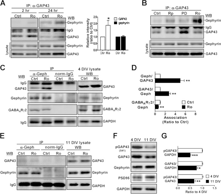
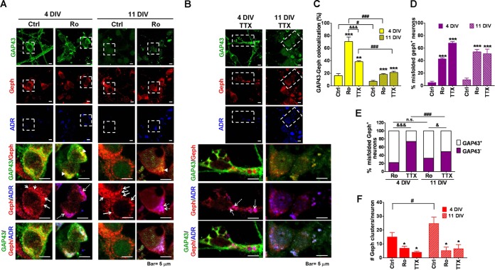
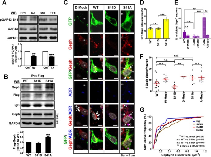
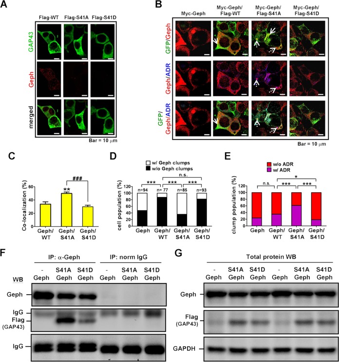
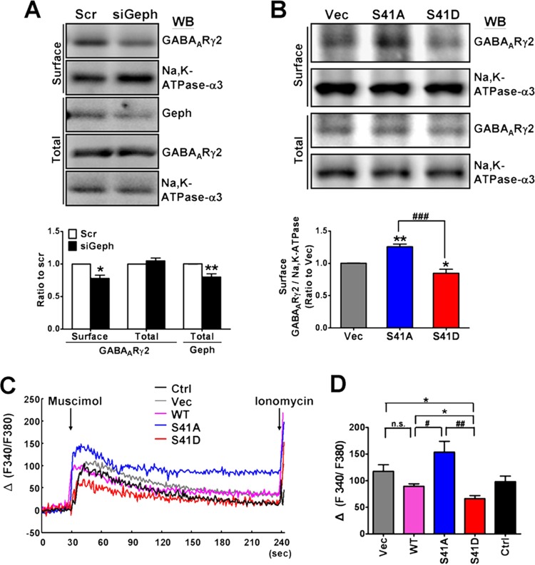
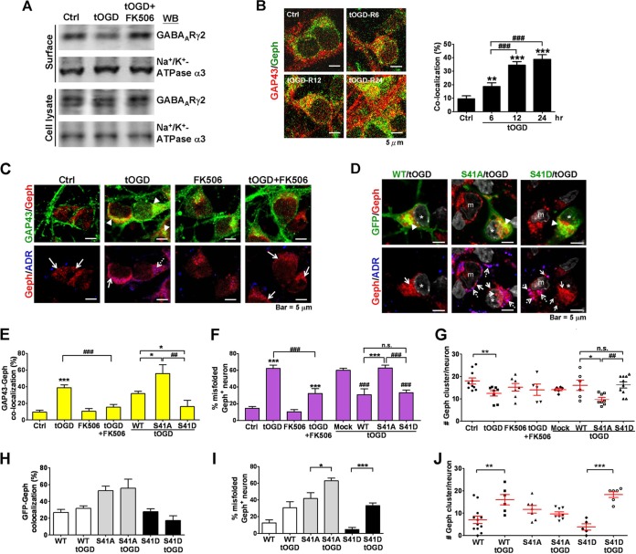
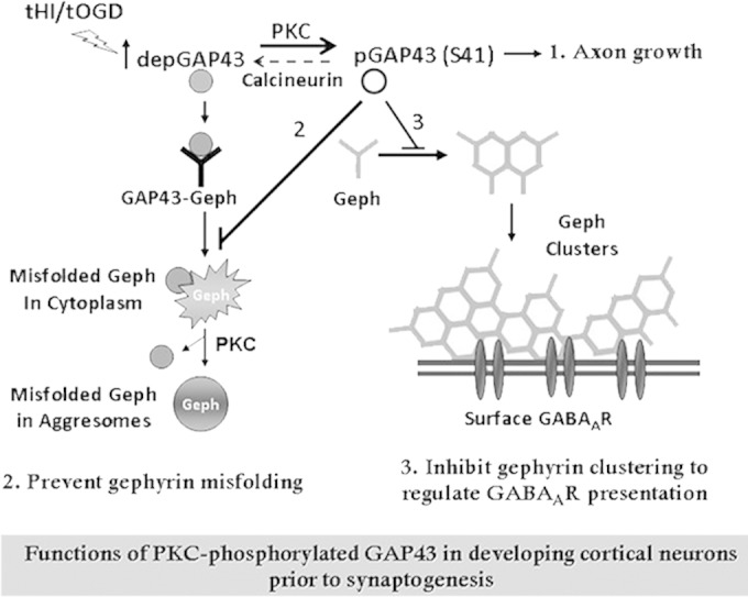
Growth-associated protein 43 (GAP43) is known to regulate axon growth, but whether it also plays a role in synaptogenesis remains unclear. Here, we found that GAP43 regulates the aggregation of gephyrin, a pivotal protein for clustering postsynaptic GABA(A) receptors (GABA(A)Rs), in developing cortical neurons. Pharmacological blockade of either protein kinase C (PKC) or neuronal activity increased both GAP43-gephyrin association and gephyrin misfolding-induced aggregation, suggesting the importance of PKC-dependent regulation of GABAergic synapses. Furthermore, we found that PKC phosphorylation-resistant GAP43(S41A), but not PKC phosphorylation-mimicking GAP43(S41D), interacted with cytosolic gephyrin to trigger gephyrin misfolding and its sequestration into aggresomes. In contrast, GAP43(S41D), but not GAP43(S41A), inhibited the physiological aggregation/clustering of gephyrin, reduced surface GABA(A)Rs under physiological conditions, and attenuated gephyrin misfolding under transient oxygen-glucose deprivation (tOGD) that mimics pathological neonatal hypoxia. Calcineurin-mediated GAP43 dephosphorylation that accompanied tOGD also led to GAP43-gephyrin association and gephyrin misfolding. Thus, PKC-dependent phosphorylation of GAP43 plays a critical role in regulating postsynaptic gephyrin aggregation in developing GABAergic synapses.


Figures

FIG 1
FIG 1
Inhibition of PKC activity induced GAP43 interaction with gephyrin in developing cortical neurons. (A) Primary rat cultured cortical neurons at 4 DIV were treated with vehicle control (Ctrl; 0.01% dimethylsulfoxide [DMSO]) or PKC inhibitor Ro318220 (Ro; 100 nM) for 2 or 24 h and then subjected to immunoprecipitation (IP) with mouse anti-GAP43 antibody (α-GAP43), followed by Western blotting (WB) with rabbit anti-GAP43 (α-GAP43) or antigephyrin (anti-Geph) antibody. Total cell lysate WB for the total level of GAP43 and gephyrin and the 24-h Ro group was quantified. (B) Neurons were treated with PKA inhibitor Rp-8-Br (Rp; 100 μM), MAPK/ERK inhibitor PD-98059 (PD; 20 μM), or Ro (100 nM) for 24 h, followed by total cell lysate WB with GAP43 and gephyrin or IP with anti-GAP43 and then WB with anti-Geph or anti-GAP43. (C and E) Immunoprecipitation with mouse anti-Geph or normal mouse IgG, followed by WB with anti-GAP43, anti-Geph, or anti-GABAARγ2 antibody in neurons at 4 (C) or 11 (E) DIV after the Ro318220 treatments. IgG served as the loading control. The total levels of GAP43, gephyrin, GABAARγ2, and GAPDH under each condition were shown in the cell lysate WB. (D) Quantification of the ratio of the two coimmunoprecipitated protein band intensities in panels A and C. (F) Differential expressions of GAP43, phosphorylated GAP43-S41 (pGAP43), gephyrin, and PSD95 in 4- and 11-DIV neurons. (G) Quantification of the relative levels of pGAP43, GAP43, and pGAP43-41/GAP43 in 4- and 11-DIV neurons. *, P < 0.05; **, P < 0.01; ***, P < 0.001 for Ctrl versus Ro in panels A and D (n = 4) and for 4 versus 11 DIV in panel G (n = 3) by unpaired t test.
FIG 2
FIG 2
Blockade of PKC or neuronal activity induced GAP43-gephyrin colocalization and gephyrin misfolding in developing cortical neurons. Immunofluorescent confocal microscopic images of 4-DIV (A) or 11-DIV (B) cortical neurons treated with vehicle (Ctrl), Ro, or TTX (1 μM) for 24 h. The merged images were from the respective dashed-line-framed area in the single-color images. Green, GAP43; red, gephyrin (Geph); blue, ADR-labeled misfolded protein. Arrowheads, GAP43-Geph colocalization (yellow signals in the GAP43/Geph panel); dashed arrows, misfolded Geph (purple signals in the Geph/ADR panel); solid arrows, red clusters in the Geph-ADR panel indicating physiological gephyrin cluster signals. (C) Quantitative results of the GAP43-Geph colocalization (n = 5 for 4 DIV; n = 15 to 35 for 11 DIV). (D) The percentage of misfolded Geph aggregate-containing neurons in each group (n = 6 for 4 DIV; n = 3 to 5 for 11 DIV). (E) The populations of misfolded Geph-containing neurons that showed GAP43+ or GAP43 immunoreactivities in the misfolded Geph aggregates under each condition. Numbers of cells counted under each condition were 91 (4 DIV-Ctrl), 119 (4 DIV-Ro), 98 (4 DIV-TTX), 77 (11 DIV-Ctrl), 46 (11 DIV-Ro), and 48 (11 DIV-TTX). n.s., not significant. (F) The number of Geph clusters per neuron in each treatment, determined by counting the red cluster signals with a diameter of >0.04 μm in the Geph-ADR merged images using MetaMorph Imaging (n = 6 for 4 DIV; n = 3 in 11 DIV). P < 0.05 (&), P < 0.01 (&&), and P < 0.001 (&&&) between Ro and TTX groups of the same DIV. P < 0.05 (#) and P < 0.001 (###) between 4 and 11 DIV of the same treatment. Fisher's exact test was used for panel E.
FIG 3
FIG 3
GAP43-S41 mutants differentially interacted with gephyrin and affected gephyrin aggregation in cortical neurons. (A) Western blotting of pGAP43-S41, total GAP43, and GAPDH in 4-DIV neurons treated with Ro (0.1 μM) or TTX (1 μM). The pGAP43-S41/GAP43 ratio (normalized by GAPDH) was used to indicate phosphorylated GAP43 at S41. **, P < 0.01; ***, P < 0.001 by unpaired t test; n = 3. (B) Coimmunoprecipitation of Flag-tagged GAP43WT (WT) or the GAP43S41A (S41A) or GAP43S41D (S41D) mutant with gephyrin (Geph) in Flag-GAP43-transfected neurons. The IgG signal served as the loading control. **, P < 0.01 versus the WT; n = 3. Total cell lysate WB of Geph showed no difference among groups. (C) Immunofluorescent confocal images of neurons transfected with GFP-tagged WT, S41D, or S41A. Green, GFP; red, Geph; blue, ADR-labeled misfolded protein; gray, DAPI. Arrowheads, GFP-Geph colocalization (yellow signals in GFP/Geph panel); arrows, physiological Geph clusters (red puncta in the Geph/ADR panel); dashed arrows, misfolded Geph (purple signals in the Geph/ADR panel); m, mock cells. (D) The percentage of GFP-Geph colocalization signals in panel C. Cell numbers counted were the following: WT, 20; S41D, 15; S41A, 18. ***, P < 0.001 versus WT results. (E) The percentage of misfolded Geph aggregate-containing neurons. W-mock, D-mock, and A-mock indicate mock cells in WT, S41D, and S41A transfection, respectively. **, P < 0.01 versus the respective mock group; P < 0.01 (##) and P < 0.001 (###) versus S41A; n = 5 to 8. (F) The number of Geph clusters per neuron. Cell numbers counted were the following: WT, 12; W-mock, 10; S41D, 5; D-mock, 5; S41A, 7; A-mock, 10. (G) Cumulative frequency of the Geph cluster area per neuron in each transfection condition. Data for the mock group were obtained from mock cells in all transfection conditions. A Kolmogorov-Smirnov test was used.
FIG 4
FIG 4
Distinct effects of GAP43 S41 mutants on gephyrin aggregation in HEK293T cell line. Immunofluorescent confocal microscopy of HEK293T cells transfected with (A) Flag-GAP43WT (Flag-WT), Flag-GAP43S41A (Flag-S41A), or Flag-GAP43S41D (Flag-S41D) or (B) Myc-Geph, Myc-Geph and Flag-WT, Myc-Geph and Flag-S41A, or Myc-Geph and Flag-S41D. Transfected cells were immunostained for Geph and GAP43. Gephyrin clumps were noted in both Myc-Geph- and Myc-Geph/Flag-S41A-transfected cells but not in the Myc-Geph/Flag-S41D-transfected cells. Dashed arrows, Geph aggregates; solid arrows, GFP-Geph colocalization. (C) GFP-Geph colocalization signals from panel B. **, P < 0.01; ###, P < 0.001; n = 15 (Geph/WT), n = 90 (Geph/S41A), and n = 88 (Geph/S41D). (D) Cell population that expressed only diffused Geph without (w/o) clumps in the cytosol or with (w/) additional Geph clumps under each condition. The number of cells counted in each group is indicated. ***, P < 0.001; n.s., not significant. (E) Population of Geph clumps colabeled with ADR or not colabeled under each condition. The numbers of Geph clumps counted in each group were the following: n = 25 (Geph), n = 20 (Geph/WT), n = 18 (Geph/S41A), and n = 16 (Geph/S41D). *, P < 0.05. (F) Co-IP of Flag-tagged GAP43 WT or S41 mutants with Geph in transfected HEK293T cells. IgG served as the loading control. (G) Western blotting of Geph, Flag, and GAPDH in the cell lysate used for panel F. Fisher's exact test was used for panels D and E.
FIG 5
FIG 5
Gephyrin knockdown and GAP43S41D overexpression reduced surface GABAAR expression. (A) Biotinylation assay of surface GABAARγ2 in gephyrin siRNA (siGeph)- and scrambled RNA (Scr)-transfected cortical neurons at 4 DIV. Na+/K+ ATPase-α3 served as the surface protein control. Total levels of GABAARγ2 and Geph were determined by cell lysate WB. P < 0.05 (*) and P < 0.01 (**) versus the Scr group by unpaired t test; n = 3. (B) Biotinylation assay of surface GABAARγ2 in cortical neurons transfected with GAP43S41A (S41A), GAP43S41D (S41D), or the vector (Vec). P < 0.05 (*) and P < 0.01 (**) versus Vec; ###, P < 0.001 between S41A and S41D groups; n = 4. (C) Representative traces and (D) compiled data of GABAAR agonist muscimol-induced [Ca2+]i increase in GFP-tagged GAP43-transfected neurons using Fura-2 calcium imaging. The calcium ionophore ionomycin (10 μM) applied at the end of each measurement confirmed the responsiveness of the observed cells. Data represent the increased ratio of Ca2+-bound to Ca2+-free Fura-2 [Δ(F340/F380)]. *, P < 0.05 versus Ctrl or Vec; P < 0.05 (#) and P < 0.01 (##) versus S41A; n = 3.
FIG 6
FIG 6
Transient hypoxia ischemia (tHI)-induced calcineurin-dependent GAP43-S41 dephosphorylation and GAP43-gephyrin association in developing neurons. Neonatal P2 rat pups were subjected to unilateral tHI or sham operation, and both contralateral (Cont) and ipsilateral (Ipsi) cerebral cortex were subjected to Western blotting of pGAP43/GAP43 in total cell lysate (A) and co-IP for GAP43-Geph association (B). (Top) representative blot; (bottom) quantification graph. The graph in panel B illustrates the data from each individual (left) and the compiled data (right). An unpaired t test was used for panel A and the compiled data in panel B (*, P < 0.05; **, P < 0.01 versus sham treatment), and paired t test was used for the individual data graph in panel B (*, P < 0.05; ns, not significant); n = 5. (C and D) Transient oxygen-glucose deprivation (tOGD), an in vitro tHI model, was applied to 4-DIV cortical neurons for 1 h, followed by 23 h of recovery under normal oxygen/glucose conditions with or without the calcineurin inhibitor FK506 (1 μM). Cell lysate WB for pGAP43-S41 and total GAP43 (C) and co-IP of GAP43 and Geph (D) were performed. ##, P < 0.01 versus tOGD; n = 4.
FIG 7
FIG 7
Calcineurin inhibitor and S41-phosphorylated GAP43 attenuated tOGD-induced GAP43-gephyrin colocalization and gephyrin misfolding. (A) Biotinylation assay of surface GABAARγ2 expression in tOGD-treated 4-DIV neurons with or without FK506 cotreatment. Na+/K+ ATPase-α3 served as a surface protein control. (B) Time-dependent increase of GAP43-gephyrin (Geph) colocalization in 4-DIV cortical neurons after tOGD. Cortical neurons were under normal conditions (Ctrl) or were subjected to 1 h of tOGD, followed by 5, 11, and 23 h of recovery (6, 12, and 24 h in total, as indicated) under normal oxygen/glucose conditions. Immunofluorescent confocal images (top) and a quantitative graph for the cocalization signals (bottom) are shown. ###, P < 0.001 versus the tOGD 6-h group; n = 6. (C) Immunofluorescent images of GAP43 (green), Geph (red), and misfolded protein aggregates (ADR; blue) in cortical neurons with or without FK506 treatment under tOGD. (D) Cortical neurons were transfected with GFP-GAP43WT (WT), GFP-GAP43S41A (S41A), or GFP-GAP43S41D (S41D), and tOGD insult was applied 24 h after the transfection. The neurons were immunolabeled with GFP (green), Geph (red), misfolded protein aggregates (ADR; blue), and cell nuclei (DAPI; gray). *, GFP-positive neurons; m, mock cell. Arrowheads in panels C and D indicate GAP43-Geph or GFP-Geph colocalization signals (yellow); dashed arrows, Geph-ADR signals (purple); solid arrows, Geph cluster signals (red clusters in Geph/ADR). (E and H) The percentage of GAP43-Geph or GFP-Geph colocalization signals; n = 5. (F and I) The population of misfolded Geph-containing neurons; n = 7. (G and J) The number of Geph clusters per neuron. Total numbers of cells counted were 164 (Ctrl), 199 (tOGD), 129 (tOGD+FK506), 6 (tOGD-mock), 6 (tOGD-WT), 8 (tOGD-S41A), and 11 (tOGD-S41D). For panels E, F, and G, P < 0.05 (*), P < 0.01 (**), and P < 0.001 (***) versus Ctrl or WT; P < 0.05 (#), P < 0.01 (##), P < 0.001 (###) versus tOGD or mock cells or between S41A and S41D. For panels H, I, and J, P < 0.05 (*), P < 0.01 (**), and P < 0.001 (***) by unpaired t test.
FIG 8
FIG 8
Proposed mode of action of GAP43 in regulating gephyrin aggregation during GABAergic synapse development. Developing cortical neurons at the presynaptogenesis stage are enriched with PKC/activity-dependent S41-phosphorylated GAP43 [pGAP43(S41)], which is known for axon growth (1). When GAP43 is dephosphorylated under physiological or pathological stimuli that inhibit PKC or activate calcineurin, dephosphorylated GAP43 (depGAP43) interacts with gephyrin (Geph) and triggers gephyrin misfolding to form cytosolic aggregates, which can be sequestered into aggresomes and then dissociated from GAP43 in a PKC-dependent manner. In contrast, pGAP43(S41) negatively regulates gephyrin clustering and surface GABAAR expression under normal development (2), yet it can effectively prevent gephyrin misfolding and facilitate gephyrin clustering under tHI insult (3). As a result, the PKC-dependent phosphorylation of GAP43 functions to regulate gephyrin-dependent surface GABAAR expression and prevent gephyrin misfolding during GABAergic synapse development.

References

    1. Alldred MJ, Mulder-Rosi J, Lingenfelter SE, Chen G, Luscher B. 2005. Distinct gamma2 subunit domains mediate clustering and synaptic function of postsynaptic GABAA receptors and gephyrin. J Neurosci 25:594–603. doi:10.1523/JNEUROSCI.4011-04.2005. - DOI - PMC - PubMed
    1. Ben-Ari Y. 2002. Excitatory actions of gaba during development: the nature of the nurture. Nat Rev Neurosci 3:728–739. doi:10.1038/nrn920. - DOI - PubMed
    1. Mohler H. 2007. Molecular regulation of cognitive functions and developmental plasticity: impact of GABAA receptors. J Neurochem 102:1–12. doi:10.1111/j.1471-4159.2007.04454.x. - DOI - PubMed
    1. Fritschy JM, Brunig I. 2003. Formation and plasticity of GABAergic synapses: physiological mechanisms and pathophysiological implications. Pharmacol Ther 98:299–323. doi:10.1016/S0163-7258(03)00037-8. - DOI - PubMed
    1. Bedford FK, Kittler JT, Muller E, Thomas P, Uren JM, Merlo D, Wisden W, Triller A, Smart TG, Moss SJ. 2001. GABA(A) receptor cell surface number and subunit stability are regulated by the ubiquitin-like protein Plic-1. Nat Neurosci 4:908–916. doi:10.1038/nn0901-908. - DOI - PubMed

Publication types

MeSH terms

LinkOut - more resources