Review
. 2013 Dec;3(4):313-20.
doi: 10.1016/j.jceh.2013.10.004. Epub 2013 Nov 27.

Neonatal hemochromatosis

Affiliations
Review

Neonatal hemochromatosis

Amy G Feldman et al. J Clin Exp Hepatol. 2013 Dec.

Abstract

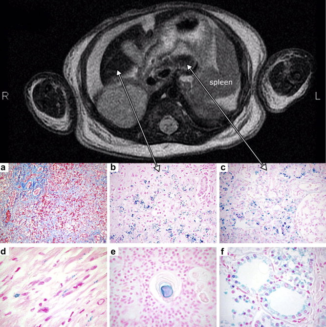
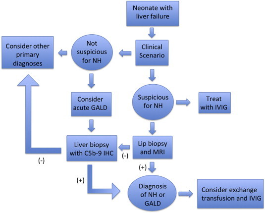
Neonatal hemochromatosis is a clinical condition in which severe liver disease in the newborn is accompanied by extrahepatic siderosis. Gestational alloimmune liver disease (GALD) has been established as the cause of fetal liver injury resulting in nearly all cases of NH. In GALD, a women is exposed to a fetal antigen that she does not recognize as "self" and subsequently begins to produce IgG antibodies that are directed against fetal hepatocytes. These antibodies bind to fetal liver antigen and activate the terminal complement cascade resulting in hepatocyte injury and death. GALD can cause congenital cirrhosis or acute liver failure with and without iron overload and siderosis. Practitioners should consider GALD in cases of fetal demise, stillbirth, and neonatal acute liver failure. Identification of infants with GALD is important as treatment is available and effective for subsequent pregnancies.

Keywords: FcRn, fragment receptor; GALD, gestational alloimmune liver disease; IVIG, intravenous immunoglobulin; IgG, immunoglobulin G; NH, neonatal hemochromatosis; NTBI, non-transferrin bound iron; acute liver failure; complement; gestational alloimmune liver disease; immunoglobulin.


Figures

Figure 1
Figure 1
MRI and histopathology of NH. Liver failure was noted at birth in a 2,900 g, 38-weeks gestation male infant. Decreased fetal movement and oligohydramnios had been noted at 36-weeks gestation. Refractory hypoglycemia and severe coagulopathy were prominent signs of liver failure. NH was suspected. On magnetic resonance imaging performed at 8 days of age, the liver (left arrow) and pancreas (right arrow) had a markedly reduced T2 signal intensity (dark area) relative to the spleen. Serum iron indices were not measured. Supportive therapy failed, and the infant expired on day of life 9. The composite image shows the most prominent histological findings on the postmortem examination. (a) Severe liver damage. A trichrome stain shows a fibrotic portal tract on the upper left. The hepatic parenchyma has been replaced by necrotic and fibrous tissue (blue-stained areas) and ductules; there were very few preserved hepatocytes (original magnification × 100). Perl's Prussian blue stain showed abnormal iron deposition in the (b) liver, (c) pancreas, (d) myocardium, (e) Hassall's corpuscles of the thymus, and (f) thyroid follicles. Original magnifications: a = 100×, b–c = 200× and d–f = 400×. The liver iron content was modestly elevated at 4,559 μg/g wet weight (reference normal 200–2,000). R, right; L, left. (Reprinted from Hepatology 2006; 43:654–660 with permission).
Figure 2
Figure 2
Diagnostic algorithm for neonatal hemochromatosis (NH). In newborns with liver failure or other clinical circumstance suspicious for NH, the infant should be given one dose of intravenous immunoglobulin (IVIG). An attempt should be made to identify extrahepatic siderosis by MRI or buccal biopsy. If either demonstrates iron overload, the diagnosis of NH is made. If both are negative, a liver biopsy for C5b-9 staining should be considered. GALD, gestational alloimmune liver disease.

References

    1. Pan X., Kelly S., Melin-Aldana H., Malladi P., Whitington P.F. Novel mechanism of fetal hepatocyte injury in congenital alloimmune hepatitis involves the terminal complement cascade. Hepatology. Jun 2010;51(6):2061–2068. - PubMed
    1. Knisely A.S., Mieli-Vergani G., Whitington P.F. Neonatal hemochromatosis. Gastroenterol Clin North Am. Sep 2003;32(3):877–889. vi–vii. - PubMed
    1. Kelly A.L., Lunt P.W., Rodrigues F. Classification and genetic features of neonatal haemochromatosis: a study of 27 affected pedigrees and molecular analysis of genes implicated in iron metabolism. J Med Genet. Sep 2001;38(9):599–610. - PMC - PubMed
    1. Hardy L., Hansen J.L., Kushner J.P., Knisely A.S. Neonatal hemochromatosis. Genetic analysis of transferrin-receptor, H-apoferritin, and L-apoferritin loci and of the human leukocyte antigen class I region. Am J Pathol. Jul 1990;137(1):149–153. - PMC - PubMed
    1. Whitington P.F., Kelly S. Outcome of pregnancies at risk for neonatal hemochromatosis is improved by treatment with high-dose intravenous immunoglobulin. Pediatrics. Jun 2008;121(6):e1615–1621. - PubMed