

Sinusoidal obstruction syndrome (hepatic veno-occlusive disease)
- PMID: 25755580
- PMCID: PMC4298625
- DOI: 10.1016/j.jceh.2014.10.002
Sinusoidal obstruction syndrome (hepatic veno-occlusive disease)
Abstract
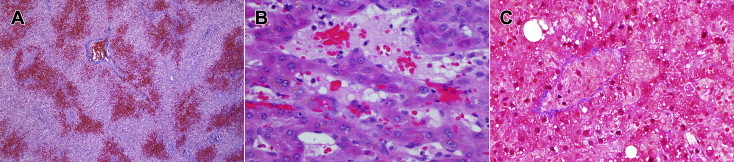
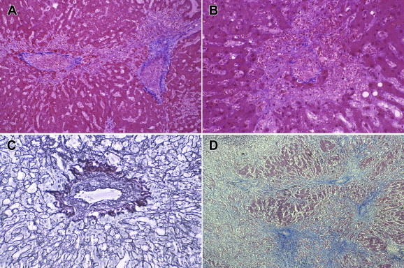
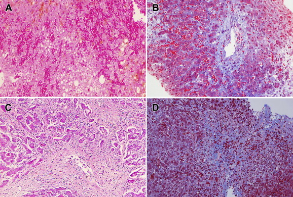
Hepatic sinusoidal obstruction syndrome (SOS) is an obliterative venulitis of the terminal hepatic venules, which in its more severe forms imparts a high risk of mortality. SOS, also known as veno-occlusive disease (VOD), occurs as a result of cytoreductive therapy prior to hematopoietic stem cell transplantation (HSCT), following oxaliplatin-containing adjuvant or neoadjuvant chemotherapy for colorectal carcinoma metastatic to the liver and treated by partial hepatectomy, in patients taking pyrrolizidine alkaloid-containing herbal remedies, and in other particular settings such as the autosomal recessive condition of veno-occlusive disease with immunodeficiency (VODI). A central pathogenic event is toxic destruction of hepatic sinusoidal endothelial cells (SEC), with sloughing and downstream occlusion of terminal hepatic venules. Contributing factors are SEC glutathione depletion, nitric oxide depletion, increased intrahepatic expression of matrix metalloproteinases and vascular endothelial growth factor (VEGF), and activation of clotting factors. The clinical presentation of SOS includes jaundice, development of right upper-quadrant pain and tender hepatomegaly, ascites, and unexplained weight gain. Owing to the potentially critical condition of these patients, transjugular biopsy may be the preferred route for liver biopsy to exclude other potential causes of liver dysfunction and to establish a diagnosis of SOS. Treatment includes rigorous fluid management so as to avoid excessive fluid overload while avoiding too rapid diuresis or pericentesis, potential use of pharmaceutics such as defibrotide, coagulolytic agents, or methylprednisolone, and liver transplantation. Proposed strategies for prevention and prophylaxis include reduced-intensity conditioning radiation for HSCT, treatment with ursodeoxycholic acid, and inclusion of bevacizumab with oxaliplatin-based chemotherapeutic regimes. While significant progress has been made in understanding the pathogenesis of SOS and in mitigating against its adverse outcomes, this condition remains a serious complication of a selective group of medical treatments.
Keywords: AML, acute myeloid leukemia; APRI, aspartate aminotransferase to platelet ratio; AST, aspartate aminotransferase; Bmab, bevacizumab; Colorectal cancer; DF, defibrotide; FOLFOX, chemotherapy regimen containing Folinic acid, 5-Fluorouracil, and Oxaliplatin; GO, gemtuzumab ozogamicin; GSTM1, glutathione S-transferase M1; GVHD, graft-versus-host disease; HSCT, hematopoietic stem cell transplantation; Hematopoietic stem cell transplantation; Herbal remedies; Liver; MOF, multi-organ failure; Oxaliplatin; PML, promyelocytic leukemia protein; RIC-HSCT, reduced-intensity conditioning hematopoietic stem cell transplantation; RILD, radiation-induced liver disease; RT, radiation therapy; SEC, sinusoidal endothelial cells; SOS, sinusoidal obstruction syndrome; TBI, total body irradiation; TIPS, transjugular intrahepatic porto-systemic shunt; UPLC-MS, ultra-performance liquid chromatography-mass spectrometry; V-PYRRO/NO, O(2)-vinyl 1-(pyrrolidin-1-yl)diazen-1-ium-1,2-diolate; VEGF, vascular endothelial growth factor; VEGFR, vascular endothelial growth factor receptor; VOD, veno-occlusive disease; VODI, veno-occlusive disease with immunodeficiency; l-NAME, N(G)-nitro-l-arginine methyl ester; s-ICAM-1, soluble intercellular adhesion molecular-1; t-PA, tissue plasminogen activator; v-WF, von Willebrand factor.
Figures
References
-
- Hess J. Fatal obliterative endophlebitis of the hepatic vein. Am J Med Sci. 1905;130:986–1001.
-
- Bras G., Jelliffe D.B., Stuart K.I. Veno-occlusive disease of liver with nonportal type of cirrhosis, occurring in Jamaica. Arch Pathol Lab Med. 1954;57:285–300. - PubMed
-
- Hahn P.F., Jackson M.A., Goldi H. Liver cirrhosis with ascites, induced in dogs by chronic massive hepatic irradiation with radioactive colloidal gold. Science. 1951;114:303–305. - PubMed
-
- Bras G., McLean E. Toxic factors in veno-occlusive disease. In: Sterling J.A., editor. Fetal and Infant Liver Function and Structure. Vol. 111. 1963. pp. 392–398. (Ann N Y Acad Sci). - PubMed
Publication types
LinkOut - more resources
Full Text Sources
Other Literature Sources
Research Materials
Miscellaneous
