. 2015 Feb 23:6:91.
doi: 10.3389/fpls.2015.00091. eCollection 2015.

Polyol specificity of recombinant Arabidopsis thaliana sorbitol dehydrogenase studied by enzyme kinetics and in silico modeling

Affiliations

Polyol specificity of recombinant Arabidopsis thaliana sorbitol dehydrogenase studied by enzyme kinetics and in silico modeling

M Francisca Aguayo et al. Front Plant Sci. .

Abstract

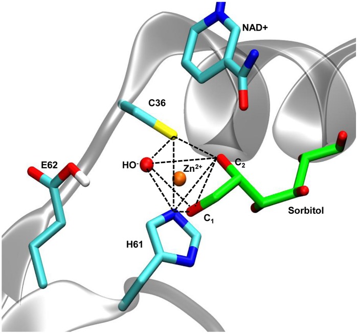
Polyols are enzymatically-produced plant compounds which can act as compatible solutes during periods of abiotic stress. Nicotinamide adenine dinucleotide(+)-dependent SORBITOL DEHYDROGENASE (SDH, E. C. 1.1.1.14) from Arabidopsis thaliana L. sorbitol dehydrogenase (AtSDH) is capable of oxidizing several polyols including sorbitol, ribitol, and xylitol. In the present study, enzymatic assays using recombinant AtSDH demonstrated a higher specificity constant for xylitol compared to sorbitol and ribitol, all of which are C2 (S) and C4 (R) polyols. Enzyme activity was reduced by preincubation with ethylenediaminetetraacetic acid, indicating a requirement for zinc ions. In humans, it has been proposed that sorbitol becomes part of a pentahedric coordination sphere of the catalytic zinc during the reaction mechanism. In order to determine the validity of this pentahedric coordination model in a plant SDH, homology modeling, and Molecular Dynamics simulations of AtSDH ternary complexes with the three polyols were performed using crystal structures of human and Bemisia argentifolii (Genn.) (Hemiptera: Aleyrodidae) SDHs as scaffolds. The results indicate that the differences in interaction with structural water molecules correlate very well with the observed enzymatic parameters, validate the proposed pentahedric coordination of the catalytic zinc ion in a plant SDH, and provide an explanation for why AtSDH shows a preference for polyols with a chirality of C2 (S) and C4 (R).

Keywords: Arabidopsis thaliana; homology modeling; molecular dynamics simulation; pentavalent zinc; polyol.


Figures

FIGURE 1
FIGURE 1
Expression and purification of recombinant His-AtSDH and AtSDH. (A) Coomassie stained gels containing purified recombinant AtSDH, before (lanes His-AtSDH) and after (lanes AtSDH) TEV-mediated cleavage of the His-tag; note the difference in migration of recombinant His-AtSDH vs. AtSDH. C+; whole reaction products from the in vitro expression of recombinant His-AtSDH. (B) Immunoblot analysis of the samples in (A), using an anti-His antisera. The dashed lines denote that this image is a reconstruction of the original immunoblot, which is shown in Supplementary Figure S1.
FIGURE 2
FIGURE 2
Kinetic properties of recombinant AtSDH. (A) Specific activities were obtained with sorbitol, ribitol, and xylitol as the variable substrate, whilst the co-substrate concentration was held saturating at 1.36 mM NAD+. The continuous lines represent the fit to a hyperbolic function. The values are the means ± SD of at least three independent determinations. (B) Specific activities were obtained with xylitol as the variable substrate, whilst the co-substrate concentration was held at 34 μM, 68 μM, or 1.36 mM NAD+. The continuous lines represent the fit to a substrate inhibition model (not shown). The values are the means ± SE of three independent determinations.
FIGURE 3
FIGURE 3
Modeling the structure of NAD+-bound AtSDH. (A) Secondary structure prediction of the N-terminus of AtSDH. The prediction was performed using Jpred3 and the secondary structure observed after modeling ab initio by loop refinement (GalaxyWeb). (B) 3D representation of a monomer of NAD+-bound AtSDH (blue ribbons) obtained by homology modeling followed by ab initio modeling of the 18 amino acid residues at the N-terminus (blue ribbon enlarged within the dashed circle). The results of the ProsaII energy and Verify3D scores for the N-terminus and the rest of the protein are shown. The structural and catalytic zinc atoms are colored orange, NAD+ is colored brown and a water molecule coordinated by the catalytic zinc atom is shown in red. (C) RMSD graph of the N-terminus (blue line) and of NAD+-bound AtSDH without the N-terminus (black line) during a 10 ns simulation. Snapshots of modeled structures of the N-terminus are shown at different time points.
FIGURE 4
FIGURE 4
Coordination geometry of the catalytic zinc of AtSDH in complex with sorbitol. In AtSDH (gray cartoon), Sγ of Cys36, N𝜀 of His61, and an oxygen atom of a water molecule form part of the trigonal bipyramidal coordination of the catalytic zinc (dashed black lines); the O1 and O2 atoms of sorbitol complete the coordination sphere. The sidechain of Glu62 was simulated in a protonated state to avoid interference with the penta coordination (see Modeling of Interactions of AtSDH Ternary Complexes with Different Polyols). The nicotinamide moiety of NAD+ is shown in close proximity to the polyol.
FIGURE 5
FIGURE 5
Hydrogen bond interactions between the AtSDH models and the polyols. The hydrogen bonds formed between AtSDH and sorbitol, ribitol, or xylitol, were quantified every 25 ps during the trajectories (left hand panels). The protein residues of the binding pocket identified in each case are represented in the right hand panels, in which dashed blue lines mark those hydrogen bonds that were maintained for at least 1 ns. The interactions with the arginine guanidinium group (R292) exhibit bidentation, hence contributing with two effective hydrogen bonds. The colors of the atoms are the same as described in Figure 3.
FIGURE 6
FIGURE 6
Water molecules present in the binding pocket of sorbitol, ribitol, and xylitol. The radial pair distribution function was calculated for the water molecules at different distances from the polyols in the active site of AtSDH (upper left panel). The remaining panels show the localization of the visually-tracked structural water molecules which mediate the interaction between the residues of AtSDH and the respective polyols. The colors of the atoms are the same as those described in Figure 3.

References

    1. Aguayo M. F., Ampuero D., Mandujano P., Parada R., Muñoz R., Gallart M., et al. (2013). Sorbitol dehydrogenase is a cytosolic protein required for sorbitol metabolism in Arabidopsis thaliana. Plant Sci. 205–206, 63–75 10.1016/j.plantsci.2013.01.012 - DOI - PubMed
    1. Alberts I., Nadassy K., Wodak S. (1998). Analysis of zinc binding sites in protein crystal structures. Protein Sci. 7 1700–1716 10.1002/pro.5560070805 - DOI - PMC - PubMed
    1. Altschul S., Gish W., Miller W., Myers E., Lipman D. (1990). Basic local alignment search tool, J. Mol. Biol. 215 403–410 10.1016/S0022-2836(05)80360-2 - DOI - PubMed
    1. Bais P., Moon S. M., He K., Leitao R., Dreher K., Walk T., et al. (2010). PlantMetabolomics.org: a web portal for plant metabolomics experiments. Plant Physiol. 152 1807–1816 10.1104/pp.109.151027 - DOI - PMC - PubMed
    1. Banfield M., Salvucci M., Baker E., Smith C. (2001). Crystal structure of the NADP(H)-dependent ketose reductase from Bemisia argentifolii at 2.3 Å resolution. J. Mol. Biol. 306 239–250 10.1006/jmbi.2000.4381 - DOI - PubMed

LinkOut - more resources