. 2015 Jan 1;8(1):441-9.
eCollection 2015.

Panax notoginseng saponins promote wound repair of anterior cruciate ligament through phosphorylation of PI3K, AKT and ERK

Affiliations

Panax notoginseng saponins promote wound repair of anterior cruciate ligament through phosphorylation of PI3K, AKT and ERK

Lu Yu et al. Int J Clin Exp Pathol. .

Abstract

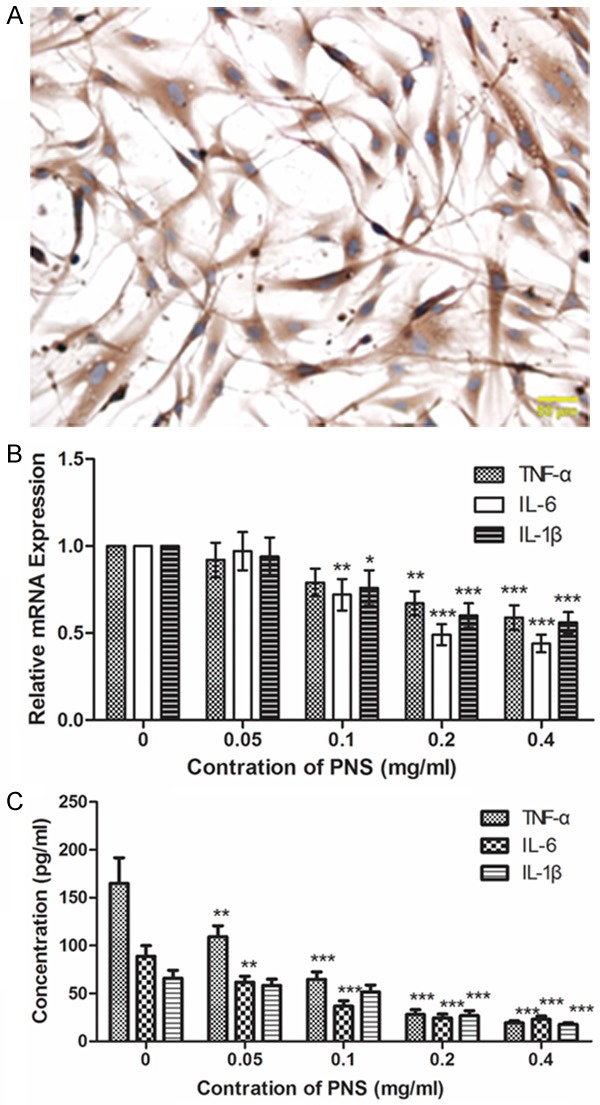
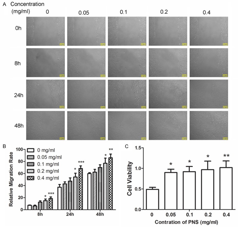
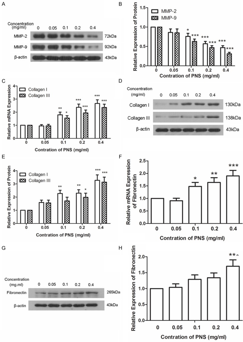
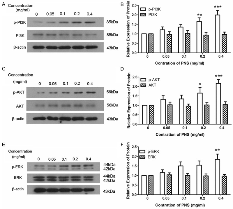
Panax notoginseng saponins (PNS) are components derived from Chinese herb panax notoginseng and play important roles in the cure of wounds. However, how PNS plays this function is still unclear. In this study, we used MTT assay, wound healing assay, western blot, quantitative real time PCR and enzyme-linked immunosorbent assay to detect the effects of PNS on the proliferation, migration and expression of collagen and fibronectin of anterior cruciate ligament (ACL) fibroblasts as well as the underlying mechanism. We found that PNS promoted the proliferation and migration of ACL fibroblasts and increased the expression levels of collagen and fibronectin. Further mechanism study indicates that PNS might play its function through the phosphorylation of PI3K, AKT and ERK. This study provides a possible mechanism for the function of PNS and lays foundation for further study on the function of panax notoginseng.

Keywords: ERK; PI3K/AKT; Panax notoginseng saponins; collagen; migration; proliferation.


Figures

Figure 1
Figure 1
PNS relieves inflammatory response in ACL fibroblasts. A. Isolated ACL fibroblasts were identified by Immunohistochemistry. Cells were stained with antibody against Vimentin. Scale bar = 50 μm. B. The mRNA expression levels of TNF-α, IL-6 and IL-1β were detected by qPCR after treatment with 0, 0.05, 0.1, 0.2 and 0.4 mg/ml of PNS. The relative mRNA expression levels were calculated using the 2-∆∆Ct method and β-actin was used as a reference. C. Concentrations of TNF-α, IL-6 and IL-1β in the media were measured by ELISA after treatment with different concentration of PNS. Each experiment was repeated three times. Results were presented as mean ± SD. *P < 0.05, **P < 0.01, ***P < 0.001.
Figure 2
Figure 2
PNS promotes the proliferation and migration of ACL fibroblasts. A, B. The migration capability of cells was detected by wound healing assay after treatment with 0, 0.05, 0.1, 0.2 and 0.4 mg/ml of PNS. C. The cell viability was detected by MTT assay after treatment with different concentration of PNS. All experiments were repeated three times and the results were presented as mean ± SD. *P < 0.05, **P < 0.01, ***P < 0.001.
Figure 3
Figure 3
PNS promotes the expression of collagen and fibronectin. A, B. Protein levels of MMP-2 and MMP-9 were measured by western blot after treatment with 0, 0.05, 0.1, 0.2 and 0.4 mg/ml of PNS. The relative expression levels of MMP-2 and MMP-9 were calculated using β-actin as reference. C. The mRNA levels of collagen I and collagen III were measured by qPCR after treatment with different concentration of PNS. The relative mRNA expression levels were calculated using the 2-∆∆Ct method with β-actin as reference. D, E. The protein levels of collagen I and collagen III were detected by western blot after treatment with different concentration of PNS. β-actin was used as reference when the relative expression levels of protein were calculated. F. The mRNA level of fibronectin was detected by qPCR using β-actin as reference. The relative mRNA expression levels were calculated using the 2-∆∆Ct method. G, H. The protein level of fibronectin was detected by western blot using β-actin as reference. All experiments were repeated three times and the results were presented as mean ± SD. *P < 0.05, **P < 0.01, ***P < 0.001.
Figure 4
Figure 4
PNS promotes the phosphorylation of PI3K, AKT and ERK. A, B. The protein levels of PI3K and p-PI3K were detected by western blot after treatment with 0, 0.05, 0.1, 0.2 and 0.4 mg/ml of PNS. The relative protein levels were calculated using β-actin as reference. C, D. The protein levels of AKT and p-AKT were measured by western blot after treatment with different concentration of PNS. β-actin was used as reference when the relative protein levels were calculated. E, F. Western blot was used to detect the protein levels of ERK and p-ERK using β-actin as reference. Each experiment was repeated three times and the results were presented as mean ± SD. *P < 0.05, **P < 0.01, ***P < 0.001.

Similar articles

Cited by

References

    1. Elsaid KA, Fleming BC, Oksendahl HL, Machan JT, Fadale PD, Hulstyn MJ, Shalvoy R, Jay GD. Decreased lubricin concentrations and markers of joint inflammation in the synovial fluid of patients with anterior cruciate ligament injury. Arthritis Rheum. 2008;58:1707–1715. - PMC - PubMed
    1. Shafizadeh S, Schneider MM, Bouillon B. Injuries of the anterior cruciate ligament in athletes. Chirurg. 2014;85:888–894. - PubMed
    1. Witonski D. [Natural history of anterior cruciate and medial collateral ligament healing--literature review] . Chir Narzadow Ruchu Ortop Pol. 2002;67:93–97. - PubMed
    1. Dong TT, Cui XM, Song ZH, Zhao KJ, Ji ZN, Lo CK, Tsim KW. Chemical assessment of roots of Panax notoginseng in China: regional and seasonal variations in its active constituents. J Agric Food Chem. 2003;51:4617–4623. - PubMed
    1. Lee MR, Yun BS, Sung CK. Comparative Study of White and Steamed Black Panax ginseng, P. quinquefolium, and P. notoginseng on Cholinesterase Inhibitory and Antioxidative Activity. J Ginseng Res. 2012;36:93–101. - PMC - PubMed

MeSH terms

Substances

LinkOut - more resources