

Progressive and painful wound as a feature of subcutaneous panniculitis-like T-cell lymphoma (SPTCL): report of a case and review of literature
- PMID: 25755768
- PMCID: PMC4348917
Progressive and painful wound as a feature of subcutaneous panniculitis-like T-cell lymphoma (SPTCL): report of a case and review of literature
Abstract
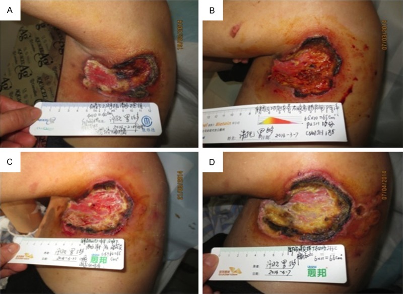
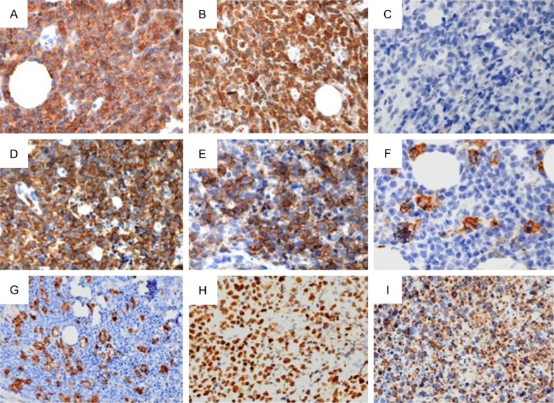
Subcutaneous panniculitis-like T-cell lymphoma (SPTCL) is an uncommon extranodal non-Hodgkin lymphoma, with an aggressive course with no well-defined treatment. This article describes a 56-year-old man, treated surgically 7 months earlier for a subcutaneous nodosity near the left axilla, presenting with a progressive inflamed wound, pain, and high fever (39 °C). Treatment with systemic antibiotics and topical anti-inflammatory dressings failed. After 7 months, the patient was diagnosed with SPTCL based on biopsy results and a multidisciplinary consultation. While undergoing systemic chemotherapy with corticosteroid therapy, his wound become more painful, larger, and covered with necrotic tissue. Fifty days after chemotherapy with corticosteroid therapy, his wound became seriously painful and increasingly necrotic. He developed a serious stomachache and abdominal distension, rapidly became comatose, and died. The aim of this case report is to present our experience of the different clinical signs of SPTCL to expedite its early diagnosis in future. We summarize the main clinical characteristics of SPTCL as a rapidly progressing and increasingly painful wound with necrotic tissue, involving a multisystem disorder, which is easily misdiagnosed, responds poorly to corticosteroid and chemotherapy treatments, and has a high mortality rate. The pathological characteristics are early inflammation, advancing to profuse infiltration of the subcutaneous adipose tissues by CD3(+) and/or CD8(+) T-cell lymphoma cells. Clinicians must cooperate with pathologists and oncologists to diagnose this disease as soon as possible and to avoid a misdiagnosis. The use of antibiotic and painkillers should minimize the patient's discomfort and control rapid wound development. Future studies are required to investigate the optimal wound treatment and whether the necrotic tissue should be removed.
Keywords: Wound; clinical characteristic; pathology; subcutaneous panniculitis T-cell lymphoma.
Figures
References
-
- Willemze R, Jaffe ES, Burg G, Cerroni L, Berti E, Swerdlow SH, Ralfkiaer E, Chimenti S, Diaz-Perez JL, Duncan LM, Grange F, Harris NL, Kempf W, Kerl H, Kurrer M, Knobler R, Pimpinelli N, Sander C, Santucci M, Sterry W, Vermeer MH, Wechsler J, Whittaker S, Meijer CJ. WHO-EORTC classification for cutaneous lymphomas. Blood. 2005;105:3768–3785. - PubMed
-
- Liau JY, Chuang SS, Chu CY, Ku WH, Tsai JH, Shih TF. The presence of clusters of plasmacytoid dendritic cells is a helpful feature for differentiating lupus panniculitis from subcutaneous panniculitis-like T-cell lymphoma. Histopathology. 2013;62:1057–1066. - PubMed
-
- Takahashi Y, Takata K, Kato S, Sato Y, Asano N, Ogino T, Hashimoto K, Tashiro Y, Takeuchi S, Masunari T, Hiramatsu Y, Maeda Y, Tanimoto M, Yoshino T. Clinicopathological analysis of 17 primary cutaneous T-cell lymphoma of the gammadelta phenotype from Japan. Cancer Sci. 2014;105:912–923. - PMC - PubMed
-
- Kim JW, Chae EJ, Park YS, Lee HJ, Hwang HJ, Lim C, Chung HW. Radiological and clinical features of subcutaneous panniculitis-like T-cell lymphoma. J Comput Assist Tomogr. 2011;35:394–401. - PubMed
Publication types
MeSH terms
Supplementary concepts
LinkOut - more resources
Full Text Sources
Research Materials