

Decidualization of intranodal endometriosis in a postmenopausal woman
- PMID: 25755813
- PMCID: PMC4348838
Decidualization of intranodal endometriosis in a postmenopausal woman
Abstract
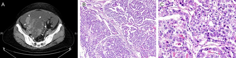
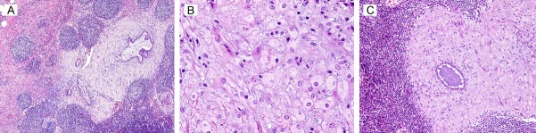
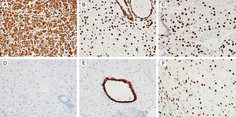
Here we describe an unusual case of decidualized endometriosis detected in pelvic lymph nodes. The presence of intranodal ectopic decidua in pregnant women has been described. A few cases of decidualization of endometriotic foci in the pelvic or para-aortic lymph nodes have also been associated with pregnancy. However, decidualized intranodal endometriosis occurring in a postmenopausal woman has not been described. A 52-year-old woman presented with a very large adnexal mass. Menopause occurred at the age of 47, and she had been treated with hormone replacement therapy. She received a total abdominal hysterectomy with bilateral salpingo-oophorectomy and pelvic and para-aortic lymphadenectomy for clear cell carcinoma of the right ovary. Histological examination revealed the presence of ectopic decidua in several pelvic lymph nodes. The deciduas consisted of sheets of loosely cohesive, large, uniform, round cells with abundant eosinophilic cytoplasm. Typical of decidualization of intranodal endometriosis, a few irregularly shaped, inactive endometrial glands lined by single layers of columnar to cuboidal epithelium were present within the decidua. An immunohistochemical study revealed that the decidual cells were positive for CD10, vimentin, estrogen receptor and progesterone receptor, which indicated that progestin-induced decidualization had occurred in the intranodal endometriotic stroma. To the best of our knowledge, this case represents the first report of decidualized intranodal endometriosis occurring in association with hormone replacement therapy in a postmenopausal woman. Misdiagnosis of this condition as a metastatic tumor can be avoided by an awareness of these benign inclusions, supported by immunohistochemical staining results.
Keywords: Decidual reaction; endometriosis; hormone replacement therapy; lymph node.
Figures
References
-
- Mills SE. Decidua and squamous metaplasia in abdominopelvic lymph nodes. Int J Gynecol Pathol. 1983;2:209–215. - PubMed
-
- Cobb CJ. Ectopic decidua and metastatic squamous carcinoma: presentation in a single pelvic lymph node. J Surg Oncol. 1988;38:126–129. - PubMed
-
- Prat J. Staging classification for cancer of the ovary, fallopian tube, and peritoneum. Int J Gynaecol Obstet. 2014;124:1–5. - PubMed
-
- Mutch DG, Prat J. 2014 FIGO staging for ovarian, fallopian tube and peritoneal cancer. Gynecol Oncol. 2014;133:401–404. - PubMed
Publication types
MeSH terms
LinkOut - more resources
Full Text Sources
Medical
Research Materials