. 2015:2015:259160.
doi: 10.1155/2015/259160. Epub 2015 Feb 12.

Differentiation between acute skin rejection in allotransplantation and T-cell mediated skin inflammation based on gene expression analysis

Affiliations

Differentiation between acute skin rejection in allotransplantation and T-cell mediated skin inflammation based on gene expression analysis

Dolores Wolfram et al. Biomed Res Int. 2015.

Abstract

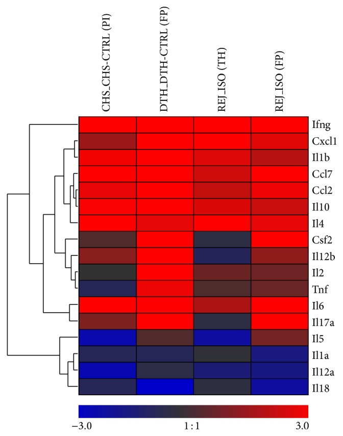
Advances in microsurgical techniques and immunosuppressive medication have rendered transplantation of vascularized composite allografts possible, when autologous tissue is neither available nor sufficient for reconstruction. However, skin rejection and side effects of long-term immunosuppression still remain a major hurdle for wide adoption of this excellent reconstructive technique. Histopathologic changes during acute skin rejection in vascular composite allotransplantation often mimic inflammatory skin disorders and are hard to distinguish. Hence, the identification of diagnostic and therapeutic markers specific for skin rejection is of particular clinical need. Here we present novel markers allowing for early differentiation between rejection in hind limb allotransplantation and contact hypersensitivity. Assessment of Ccl7, Il18, and Il1b expression is most indicative of distinguishing skin rejection from skin inflammatory disorders. Gene expression levels varied significantly across skin types and regions, indicating localization specific mechanism of leukocyte migration and infiltration. Expression of Il12b, Il17a, and Il1b gene expression levels differed significantly between rejection and inflammation, independent of the skin type. In synopsis of the RNA expression profile and previously assessed protein expression, the Il1 family appears as a promising option for accurate skin rejection diagnosis and, as a following step, for development of novel rejection treatments.


Figures

Figure 1
Figure 1
Clinical evaluation of animals. Representative pictures of skin inflammation: (a) transplanted limb showing clinically grade 2 rejection characterized by erythema and swelling; (b, c) DTH reaction on the planta pedis on the right paw and the control footpad on the left side without any inflammatory reaction.
Figure 2
Figure 2
Histological evaluation of skin samples. Representative microscopic images of hematoxylin and eosin stained histological skin sections: (a) contact hypersensitivity (CHS) reaction (pinna, 24 h), (A) control skin from left pinna, (b) skin rejection (grade 2, thigh, POD 5); (c) delayed type hypersensitivity (DTH) reaction (planta pedis, 24 h), (C) control skin from left footpad, and (d) skin rejection (grades 1-2, planta pedis) are presented.
Figure 3
Figure 3
Relative expression of 17 cytokines/chemokines mRNA extracted from samples of an inflammatory disease model (DTH, CHS) or from planta pedis or thigh in a rat hind limb transplantation model on POD 5. Relative expression levels are presented as mean + standard error of mean and normalized to the mean level in the inflammatory model.
Figure 4
Figure 4
Separation of the samples within the two combined studied groups by linear discrimination analysis (LDA). (a) Coefficients of the discriminant vector from a multivariate model indicating the contribution to the separation for each of the 17 studied genes, (b) separation of the two groups by values of the canonical variable for each sample resulting from LDA including all 17 cytokines/chemokines relative mRNA levels (ΔCT) and corresponding density distribution, (c) list of significant LDA models when studying all pairwise gene models, and (d) linear separation based on the ΔCT levels of Ccl7 and Il1b are shown. Allografts skin samples are shown in gray, inflammatory skin disease is shown in black, and dashed gray line indicates linear separation and partition.
Figure 5
Figure 5
Heat map visualizing mean log2-fold changes of cytokine/chemokine mRNA levels in two different inflammatory models (DTH, CHS) and two different skin types of allograft rejection at POD 5 in rat hind limb transplantation. Relative expressions are normalized to the mean of the controls from the respective group. Greater levels of inflammatory mediators (log2-fold changes > 0) are indicated in red and smaller levels (log2-fold changes < 0) are indicated in blue according to the legend. Similar profiles of mediators across all conditions are grouped by average linkage hierarchical clustering as indicated by the dendrogram (tree) at the left.
Figure 6
Figure 6
Distribution of log2-fold gene expression changes for 3 selected mediators (Il12b, Il1b, and Il17a) in the two inflammatory models (CHS, DTH) as well as in footpad (FP) and thigh (TH) samples from allograft rejection in rat hind limb transplantation. Relative expression levels are normalized to the mean of the controls in the respective group. Differences of the relative expressions (log2-fold changes) were tested between the rejection and the inflammatory model in the same skin type (hairy or nonhairy skin) by Tukey HSD post hoc procedure and resulting adjusted P values are provided.

Similar articles

Cited by

References

    1. Cendales L. C., Kanitakis J., Schneeberger S., et al. The Banff 2007 working classification of skin-containing composite tissue allograft pathology. The American Journal of Transplantation. 2008;8(7):1396–1400. doi: 10.1111/j.1600-6143.2008.02243.x. - DOI - PubMed
    1. Kanitakis J. The challenge of dermatopathological diagnosis of composite tissue allograft rejection: a review. Journal of Cutaneous Pathology. 2008;35(8):738–744. doi: 10.1111/j.1600-0560.2007.00889.x. - DOI - PubMed
    1. Saint-Mezard P., Berard F., Dubois B., Kaiserlian D., Nicolas J. F. The role of CD4+ and CD8+ T cells in contact hypersensitivity and allergic contact dermatitis. European Journal of Dermatology. 2004;14(3):131–138. - PubMed
    1. Pastore S., Mascia F., Mariotti F., Dattilo C., Girolomoni G. Chemokine networks in inflammatory skin diseases. European Journal of Dermatology. 2004;14(4):203–208. - PubMed
    1. Grabbe S., Schwarz T. Immunoregulatory mechanisms involved in elicitation of allergic contact hypersensitivity. Immunology Today. 1998;19(1):37–44. doi: 10.1016/S0167-5699(97)01186-9. - DOI - PubMed

MeSH terms

LinkOut - more resources