

Pachychoroid diseases of the macula
- PMID: 25756060
- PMCID: PMC4352204
Pachychoroid diseases of the macula
Abstract
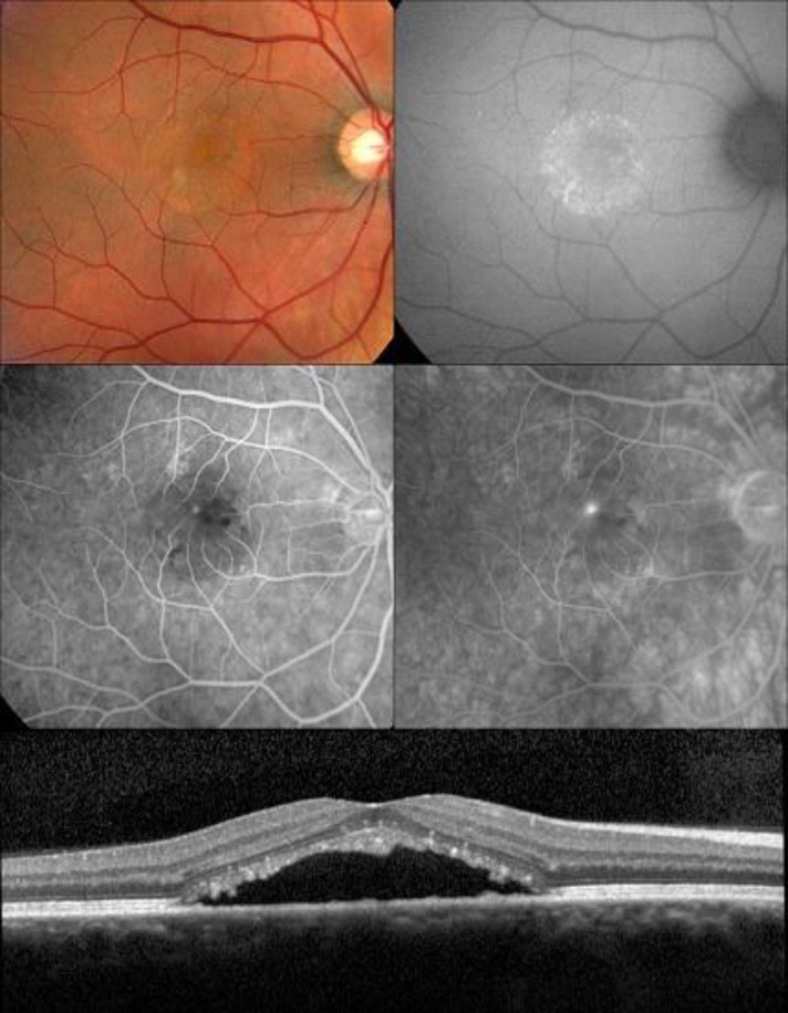
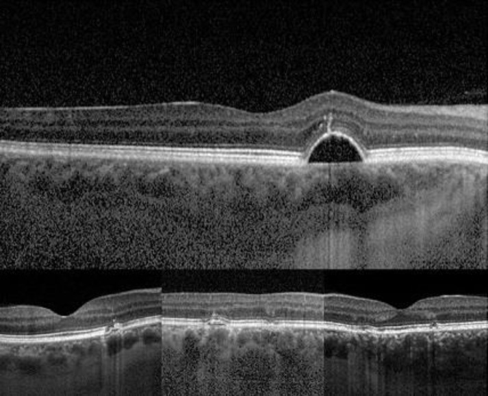
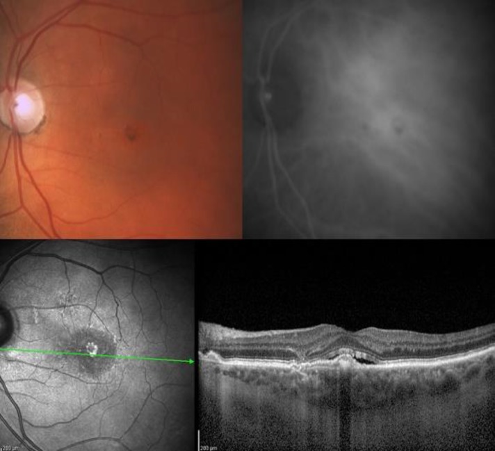
Advances in optical coherence tomography have enabled a better appreciation of the role of pathologic choroidal changes in a variety of retinal disease. A "pachychoroid" (pachy-[prefix]: thick) is defined as an abnormal and permanent increase in choroidal thickness often showing dilated choroidal vessels and other structural alterations of the normal choroidal architecture. Central serous chorioretinopathy is just one of several pachychoroid-related macular disorders. This review summarizes the current state of knowledge of the pachycoroid spectrum and the hallmark features seen with multimodal imaging analysis of these entities.
Keywords: Central serous chorioretinopathy; Macula; Pachychoroid Diseases.
Figures
References
-
- Swanson EA, Izatt JA, Hee MR, Huang D, Lin CP, Schuman JS, Puliafito CA, Fujimoto JG. In vivo retinal imaging by optical coherence tomography. Opt Lett. 1993 Nov;18(21):1864–6. PMID: 19829430. - PubMed
-
- Toth CA, Narayan DG, Boppart SA, Hee MR, Fujimoto JG, Birngruber R, Cain CP, DiCarlo CD, Roach WP. A comparison of retinal morphology viewed by optical coherence tomography and by light microscopy. Arch Ophthalmol. 1997 Nov;115(11):1425–8. PMID: 9366674. - PubMed
-
- Gloesmann M, Hermann B, Schubert C, Sattmann H, Ahnelt PK, Drexler W. Histologic correlation of pig retina radial stratification with ultrahigh-resolution optical coherence tomography. Invest Ophthalmol Vis Sci. 2003 Apr;44(4):1696–703. PMID: 12657611. - PubMed
Publication types
LinkOut - more resources
Full Text Sources