. 2015 Mar 10;10(3):e0115830.
doi: 10.1371/journal.pone.0115830. eCollection 2015.

Evaluation of the synuclein-γ (SNCG) gene as a PPARγ target in murine adipocytes, dorsal root ganglia somatosensory neurons, and human adipose tissue

Affiliations

Evaluation of the synuclein-γ (SNCG) gene as a PPARγ target in murine adipocytes, dorsal root ganglia somatosensory neurons, and human adipose tissue

Tamara N Dunn et al. PLoS One. .

Abstract

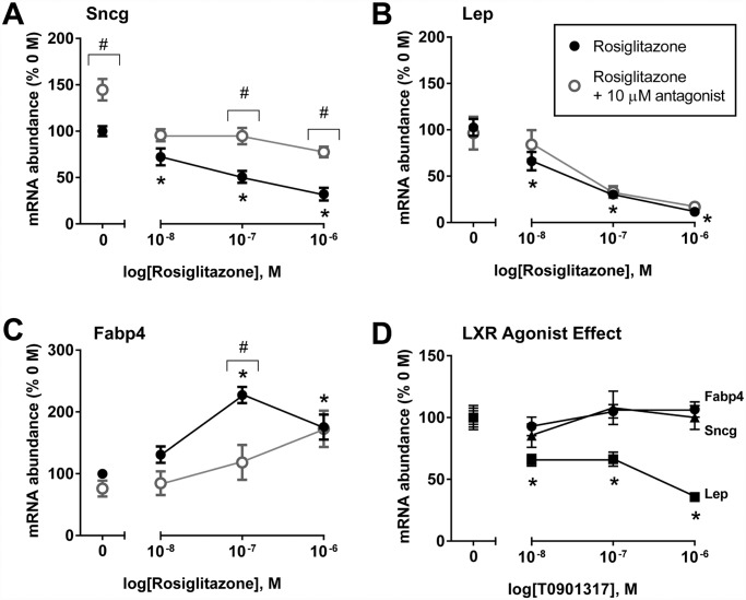
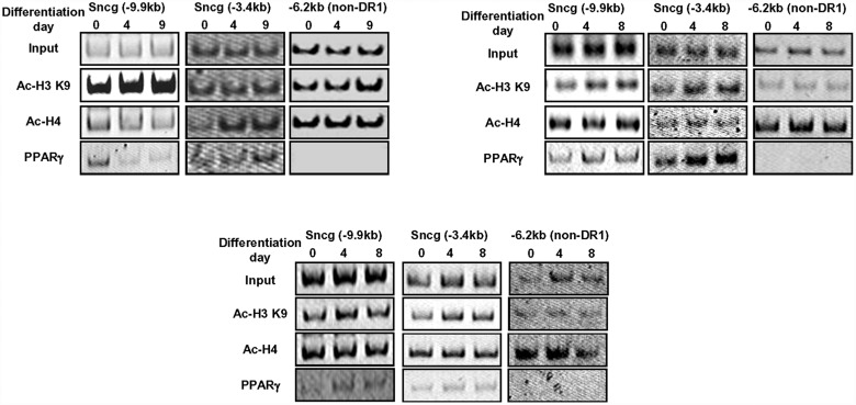
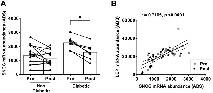
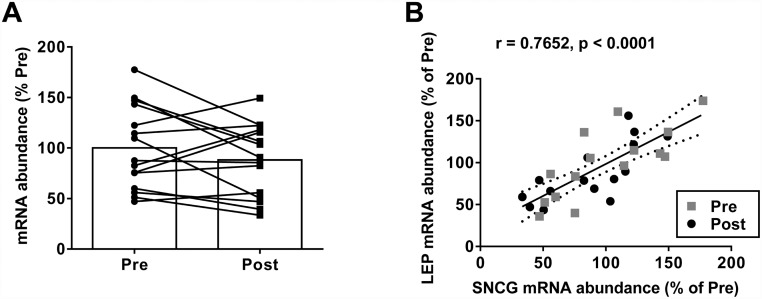
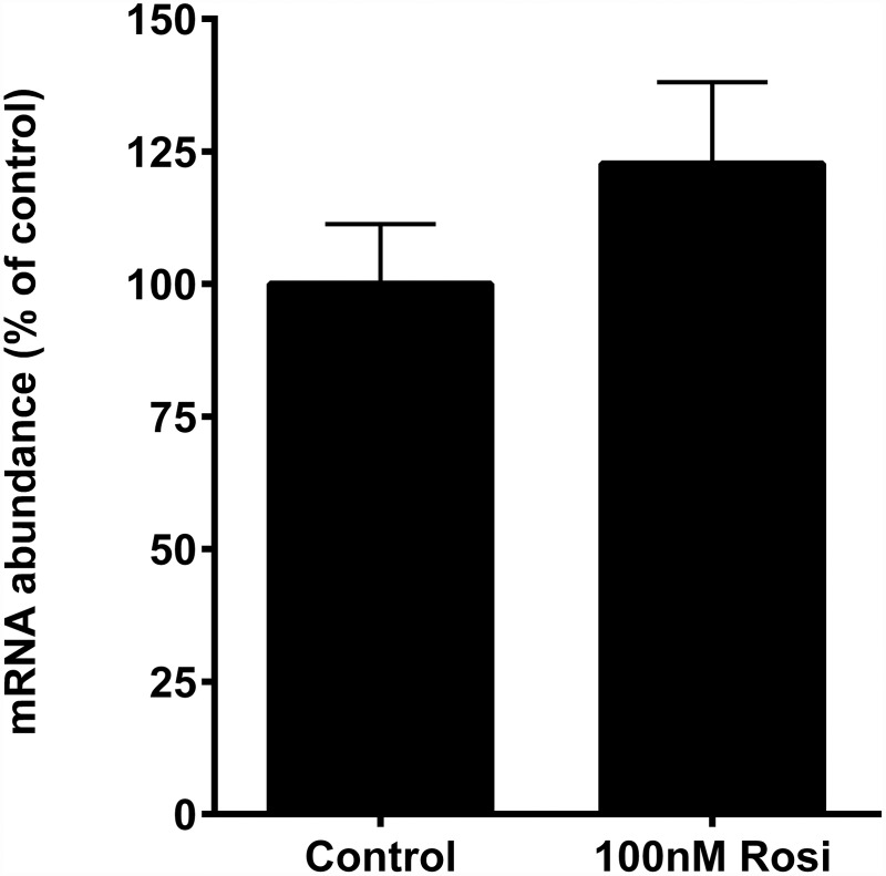
Recent evidence in adipocytes points to a role for synuclein-γ in metabolism and lipid droplet dynamics, but interestingly this factor is also robustly expressed in peripheral neurons. Specific regulation of the synuclein-γ gene (Sncg) by PPARγ requires further evaluation, especially in peripheral neurons, prompting us to test if Sncg is a bona fide PPARγ target in murine adipocytes and peripheral somatosensory neurons derived from the dorsal root ganglia (DRG). Sncg mRNA was decreased in 3T3-L1 adipocytes (~68%) by rosiglitazone, and this effect was diminished by the PPARγ antagonist T0070907. Chromatin immunoprecipitation experiments confirmed PPARγ protein binding at two promoter sequences of Sncg during 3T3-L1 adipogenesis. Rosiglitazone did not affect Sncg mRNA expression in murine cultured DRG neurons. In subcutaneous human WAT samples from two cohorts treated with pioglitazone (>11 wks), SNCG mRNA expression was reduced, albeit highly variable and most evident in type 2 diabetes. Leptin (Lep) expression, thought to be coordinately-regulated with Sncg based on correlations in human adipose tissue, was also reduced in 3T3-L1 adipocytes by rosiglitazone. However, Lep was unaffected by PPARγ antagonist, and the LXR agonist T0901317 significantly reduced Lep expression (~64%) while not impacting Sncg. The results support the concept that synuclein-γ shares some, but not all, gene regulators with leptin and is a PPARγ target in adipocytes but not DRG neurons. Regulation of synuclein-γ by cues such as PPARγ agonism in adipocytes is logical based on recent evidence for an important role for synuclein-γ in the maintenance and dynamics of adipocyte lipid droplets.


Conflict of interest statement

Competing Interests: Dr. Smith has received funding from, and served as a consultant to, Takeda Pharmaceuticals. This does not alter the authors’ adherence to PLOS ONE policies on sharing data and materials.

Figures

Fig 1
Fig 1. Regulation of mRNA expression levels for synuclein-γ (Sncg) (A), leptin (Lep) (B), and fatty acid binding protein (Fabp4) (C) by 24 hr short term treatment with the PPARγ agonist rosiglitazone, or co-treatment with PPARγ antagonist, T0070907, in mature 3T3-L1 adipocytes.
Also shown are mRNA expression levels following treatment with LXR agonist, T0901317, in mature 3T3-L1 adipocytes (D). Values are means +/- SEM, n = 7–15 per concentration; data from two experiments. Transcript level in vehicle-treated (0 M) control cells was considered 100%; gene expression values were calculated within experiment relative to control. *P<0.05, 1-way ANOVA with a post hoc Dunnett’s test comparing rosiglitazone concentrations to the vehicle-treated control. #P<0.05, Student’s t test for effect of 10 μM T0070907 antagonist at each individual concentration of rosiglitazone. Raw data is provided in S1 Data.
Fig 2
Fig 2. Temporal changes in PPARγ binding at predicted DR1 sites in the promoter region of the murine Sncg gene during 3T3-L1 adipocyte differentiation.
ChIP studies were performed in pre-adipocytes and maturing adipocytes at days 4 and 8 post-differentiation initiation, employing anti-PPARγ antibody and sequence-specific primers for putative DR1 sites (PPAR-response elements) located at promoter regions—9.9 and—3.4kb, and a non-DR1 site at-6.2 kb, relative to the murine Sncg start codon. Also shown are histone acetylation patterns at each site, employing anti-acetylated histone-3 K9 (Ac-H3 K9) and anti-acetylated histone-4 pan (Ac-H4). Images are results from 3 independent experiments. Amplicon sizes are described in the text, and a representative image showing DNA band sizes is provided as S1 Fig.
Fig 3
Fig 3. COHORT 1: 12 weeks PIO treatment significantly decreased synuclein-γ (SNCG) mRNA expression in subcutaneous white adipose tissue (SC-WAT) from pre-treatment levels in subjects with type 2 diabetes (n = 8) but not in non-diabetics (n = 17).
(A). *P<0.05, paired t test. Pre- and post-treatment SC-WAT expression levels of SNCG and LEP in the same subjects were highly-correlated in T2DM and non-diabetic subjects, using Pearson’s correlation statistic (B). ADS—average difference score calculated using Affymetrix MAS5.0. Raw data is provided in S1 Data.
Fig 4
Fig 4. COHORT 2: Effect of 11 weeks PIO treatment on synuclein-γ (SNCG) mRNA expression in subcutaneous white adipose tissue (SC-WAT) in subjects with type 2 diabetes (n = 15).
(A). Pre- and post-treatment SC-WAT expression levels of SNCG and LEP in the same subjects were highly-correlated, using Pearson’s correlation statistic (B). The average transcript level in pre-treatment biopsies was considered 100%. Raw data is provided in S1 Data.
Fig 5
Fig 5. Effect of 24 hr treatment with 100 nM PPARγ agonist, rosiglitazone (Rosi), on mRNA expression levels for synuclein-γ (Sncg) in murine primary dorsal root ganglia neurons.
Values are means +/- SEM, n = 17/treatment. Data are from four independent experiments. Transcript level in vehicle-treated control cells was considered 100%. Raw data is provided in S1 Data.

Similar articles

Cited by

References

    1. Song CK, Schwartz GJ, Bartness TJ (2009) Anterograde transneuronal viral tract tracing reveals central sensory circuits from white adipose tissue. Am J Physiol Regul Integr Comp Physiol 296: R501–511. 10.1152/ajpregu.90786.2008 - DOI - PMC - PubMed
    1. Bartness TJ, Shrestha YB, Vaughan CH, Schwartz GJ, Song CK (2010) Sensory and sympathetic nervous system control of white adipose tissue lipolysis. Molecular and cellular endocrinology 318: 34–43. 10.1016/j.mce.2009.08.031 - DOI - PMC - PubMed
    1. Bartness TJ, Vaughan CH, Song CK (2010) Sympathetic and sensory innervation of brown adipose tissue. Int J Obes (Lond) 34 Suppl 1: S36–42. - PMC - PubMed
    1. Vaughan CH, Shrestha YB, Bartness TJ (2011) Characterization of a novel melanocortin receptor-containing node in the SNS outflow circuitry to brown adipose tissue involved in thermogenesis. Brain research 1411: 17–27. 10.1016/j.brainres.2011.07.003 - DOI - PMC - PubMed
    1. van Baak MA (2001) The peripheral sympathetic nervous system in human obesity. Obes Rev 2: 3–14. - PubMed

Publication types

MeSH terms