. 2015 Jul;135(7):1849-1862.
doi: 10.1038/jid.2015.95. Epub 2015 Mar 10.

Melanoma Cell Galectin-1 Ligands Functionally Correlate with Malignant Potential

Affiliations

Melanoma Cell Galectin-1 Ligands Functionally Correlate with Malignant Potential

Erika M Yazawa et al. J Invest Dermatol. 2015 Jul.

Abstract

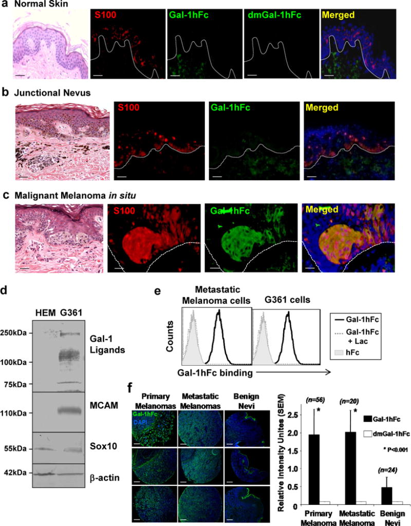
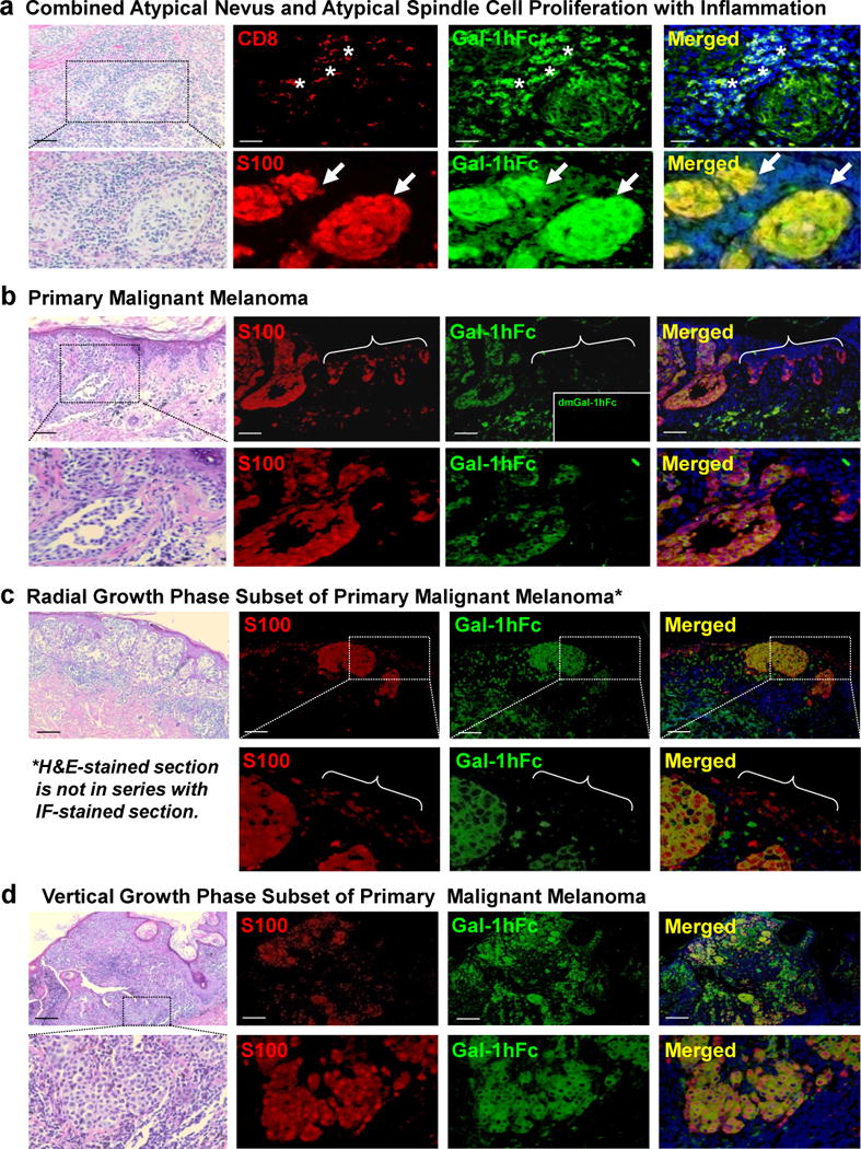
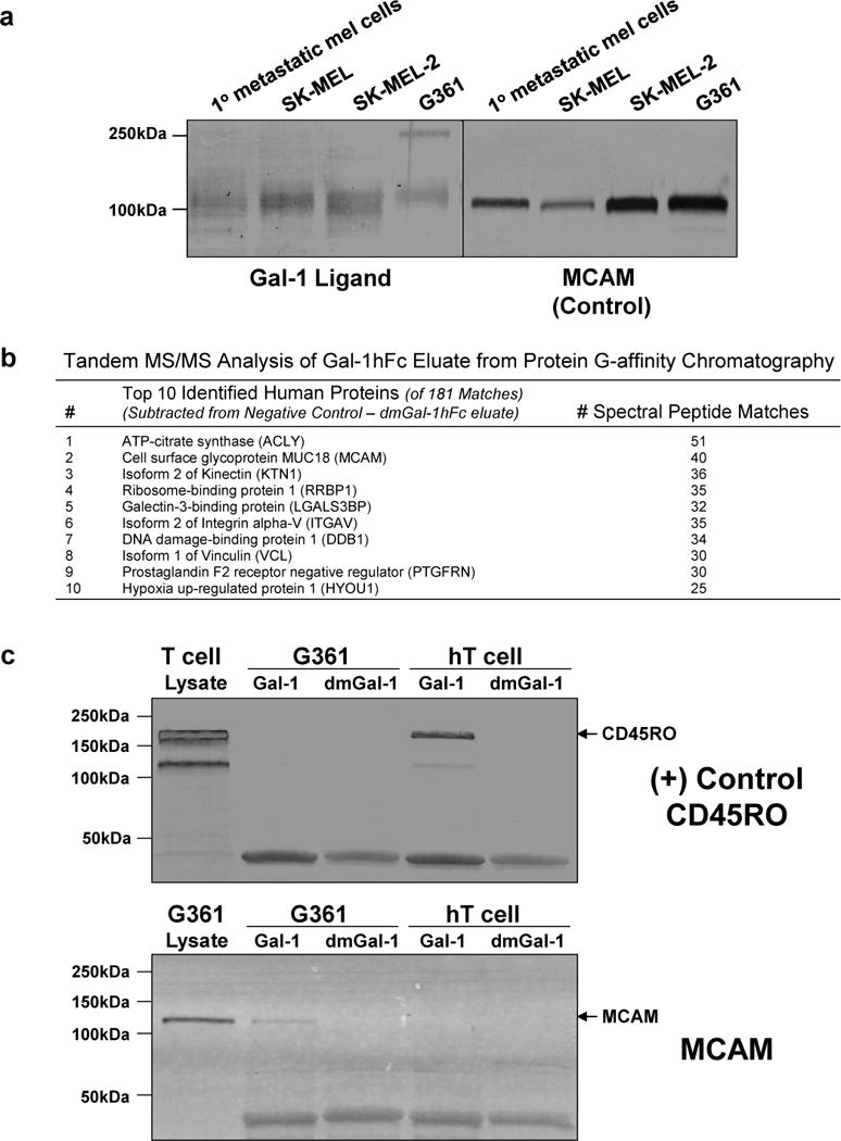
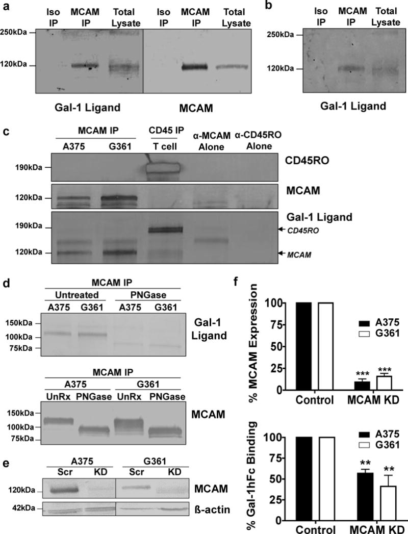
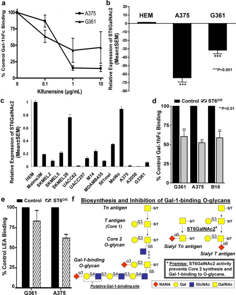
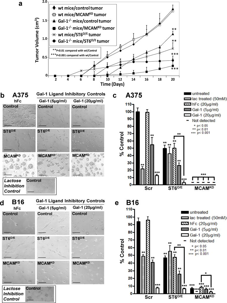
Galectin-1 (Gal-1)-binding to Gal-1 ligands on immune and endothelial cells can influence melanoma development through dampening antitumor immune responses and promoting angiogenesis. However, whether Gal-1 ligands are functionally expressed on melanoma cells to help control intrinsic malignant features remains poorly understood. Here, we analyzed expression, identity, and function of Gal-1 ligands in melanoma progression. Immunofluorescent analysis of benign and malignant human melanocytic neoplasms revealed that Gal-1 ligands were abundant in severely dysplastic nevi, as well as in primary and metastatic melanomas. Biochemical assessments indicated that melanoma cell adhesion molecule (MCAM) was a major Gal-1 ligand on melanoma cells that was largely dependent on its N-glycans. Other melanoma cell Gal-1 ligand activity conferred by O-glycans was negatively regulated by α2,6 sialyltransferase ST6GalNAc2. In Gal-1-deficient mice, MCAM-silenced (MCAM(KD)) or ST6GalNAc2-overexpressing (ST6(O/E)) melanoma cells exhibited slower growth rates, underscoring a key role for melanoma cell Gal-1 ligands and host Gal-1 in melanoma growth. Further analysis of MCAM(KD) or ST6(O/E) melanoma cells in cell migration assays indicated that Gal-1 ligand-dependent melanoma cell migration was severely inhibited. These findings provide a refined perspective on Gal-1/melanoma cell Gal-1 ligand interactions as contributors to melanoma malignancy.


Conflict of interest statement

Conflict of Interest

While all other authors declare no conflicts of interest, Dr. Abrar A. Qureshi consults for Abbvie, Inc., Amgen, Inc., Novartis Corp, the Centers for Disease Control and Prevention, Janssen, Inc. and Merck & Co., Inc., and serves as an investigator for Amgen, Inc.

Figures

Figure 1
Figure 1. Gal-1 ligands are differentially expressed on normal human melanocytes and human melanoma cells
Dual IF analysis of Gal-1 ligands with Gal-1hFc (in green) or dmGal1-hFc control and S100 with anti-S100A-B (in red) was performed on FFPE-sections of normal human skin (a), a benign junctional nevus (b) and melanoma in situ (c). In (d), Western blot analysis of Gal-1 ligands (with Gal-1hFc), MCAM polypeptide, SOX10 and ß-actin in HEM and G361 melanoma cell lysates was performed. Primary melanoma cells and G361 melanoma cells were FACS analyzed with Gal-1hFc or controls (e). In (f), IF analysis of Gal-1 ligands was performed on TMAs containing primary (n=56) and metastatic melanomas (n=20) and benign nevi (n=24). (*p<0.001; Statistically significance compared with benign nevi). Scale bars = 100μm.
Figure 2
Figure 2. A pre-malignant melanocytic tumor and malignant melanomas are strongly positive for Gal-1 ligands
Dual IF analysis of Gal-1 ligands (in green) and CD8 or S100 (in red) was performed on FFPE-sections of (a) a combined atypical nevus and an atypical spindle cell proliferation with inflammation (Asterisks= Gal-1 ligand+ CD8+ cells) (Arrows= Gal-1 ligand+ S100+ dermal nests). Dual IF staining of primary cutaneous melanomas (b), including radial (c) and vertical (d) growth phase subsets, was also performed. Brackets in (b) (Upper Panel) and (c) (Lower Panel) indicated margin tissue where non-malignant epidermal S100+ cells were Gal-1 ligand. Scale bars = 100μm and photomicrographs enlarged in the lower panels as indicated.
Figure 3
Figure 3. Affinity-purification of candidate Gal-1 ligands from human melanoma cells implicates MCAM as a major Gal-1 ligand
In (a), primary metastatic melanoma cells or melanoma cell line lysates were blotted with Gal-1hFc or control anti-MCAM. As shown in (b), the top 10 proteins and corresponding number of peptide matches identified by tandem MS/MS of elutes from protein G affinity chromatography with Gal-1hFc or negative control dmGal-1hFc and G361 cell lysate are listed. In (c), control activated human T cell or melanoma G361 cell lysate and eluates from protein G affinity chromatography with Gal-1hFc or negative control dmGal-1hFc were blotted with anti-CD45RO or anti-MCAM. Arrows indicate the presence of T cell CD45RO at 190kDa and melanoma cell MCAM at 120kD.
Figure 4
Figure 4. N-glycosylated MCAM binds Gal-1 and is a major contributor of total melanoma cell ligand activity
Anti-MCAM immunoprecipitates from G361 (a) or primary melanoma cell lysates (b) were blotted with Gal-1hFc or anti-MCAM. In (c), anti-CD45RO immunoprecipitate from activated T cell lysate or anti-MCAM immunoprecipitate from A375 and G316 cell lysates were blotted with anti-CD45RO, anti-MCAM or Gal-1hFc. Anti-MCAM immunoprecipitate from A375 and G361 cell lysates were treated with PNGase and blotted with Gal-1hFc or anti-MCAM (d). Scr or MCAMKD A375 and G361 cell lysates were blotted with anti-MCAM or anti-ß-actin (e). In (f), Scr or MCAMKD A375 and G361 cells were analyzed for MCAM and Gal-1 ligand by flow cytometry (**p<0.01 and ***p<0.001; statistically significance compared with Scr control). All experiments were performed three-times.
Figure 5
Figure 5. ST6GalNAc2 is downregulated in melanoma cells and is a putative regulator of O-glycan-dependent Gal-1 ligand activity
Kifunensine-treated A375 and G361 cells were assayed for Gal-1 ligand activity by flow cytometry with Gal-1hFc or controls (a). Real-time RT-qPCR analysis was performed on HEM, G361 and A375 cells (b) and 11 other melanoma cell lines (c). Relative ST6GalNAc2 expression level was normalized to expression in HEM over 3-experiments and expressed as Mean±SEM. Gal-1hFc- (d) and LEA-binding (e) of control or ST6O/E melanoma cells were analyzed by flow cytometry. In (f), an illustration of ST6GalNAc2’s role on the biosynthesis and putative inhibition of Gal-1-binding to O-glycans. **p<0.01 and ***p<0.001 – statistical significance compared with untreated controls or HEM.
Figure 6
Figure 6. In vivo growth of melanoma cells and migration of melanoma cells on Matrigel is regulated, in part, by host Gal-1 and on melanoma cell Gal-1 ligands
Wt or Gal-1−/− mice were inoculated s.c. with control, ST6O/E or MCAMKD B16 cells and monitored for tumor growth. Mean tumor volumes (SEM) (n=8/group) were calculated and plotted against time (a). In (b–e), control, ST6O/E or MCAMKD A375 and B16 melanoma cells pre-blocked with Gal-1hFc, hFc or lactose were assayed for formation of tube-like structures on Matrigel. Tube-like structures were illustrated in representative phase photomicrographs (Scale bars=100μm). The number of tube-like structures was expressed as % Control hFc-treated cells (*p<0.05, **p<0.01 and ***p<0.001; statistically significance compared with hFc-control cells). Data were collected from at least 3-experiments.

Similar articles

Cited by

References

    1. Aisner DL, Maker A, Rosenberg SA, et al. Loss of S100 antigenicity in metastatic melanoma. Human pathology. 2005;36:1016–9. - PMC - PubMed
    1. Andre S, Kojima S, Yamazaki N, et al. Galectins-1 and -3 and their ligands in tumor biology. Non-uniform properties in cell-surface presentation and modulation of adhesion to matrix glycoproteins for various tumor cell lines, in biodistribution of free and liposome-bound galectins and in their expression by breast and colorectal carcinomas with/without metastatic propensity. Journal of cancer research and clinical oncology. 1999;125:461–74. - PubMed
    1. Banh A, Zhang J, Cao H, et al. Tumor galectin-1 mediates tumor growth and metastasis through regulation of T-cell apoptosis. Cancer research. 2011;71:4423–31. - PMC - PubMed
    1. Barthel SR, Antonopoulos A, Cedeno-Laurent F, et al. Peracetylated 4-fluoro-glucosamine reduces the content and repertoire of N- and O-glycans without direct incorporation. The Journal of biological chemistry. 2011;286:21717–31. - PMC - PubMed
    1. Baum LG, Seilhamer JJ, Pang M, et al. Synthesis of an endogeneous lectin, galectin-1, by human endothelial cells is up-regulated by endothelial cell activation. Glycoconjugate journal. 1995;12:63–8. - PubMed

Publication types

MeSH terms

LinkOut - more resources