. 2015 Mar 10;9(3):e0003601.
doi: 10.1371/journal.pntd.0003601. eCollection 2015 Mar.

Exposure to Leishmania braziliensis triggers neutrophil activation and apoptosis

Affiliations

Exposure to Leishmania braziliensis triggers neutrophil activation and apoptosis

Sarah A C Falcão et al. PLoS Negl Trop Dis. .

Abstract

Background: Neutrophils are the first line of defense against invading pathogens and are rapidly recruited to the sites of Leishmania inoculation. During Leishmania braziliensis infection, depletion of inflammatory cells significantly increases the parasite load whereas co-inoculation of neutrophils plus L. braziliensis had an opposite effect. Moreover, the co-culture of infected macrophages and neutrophils also induced parasite killing leading us to ask how neutrophils alone respond to an L. braziliensis exposure. Herein we focused on understanding the interaction between neutrophils and L. braziliensis, exploring cell activation and apoptotic fate.

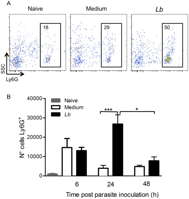
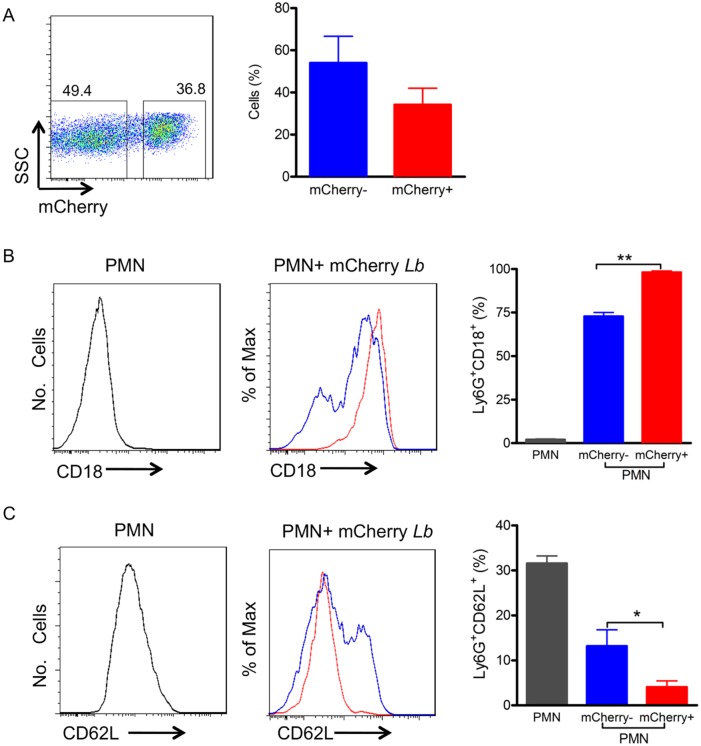
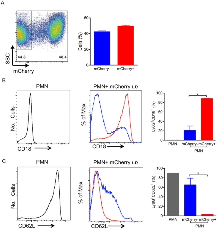
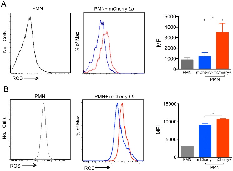
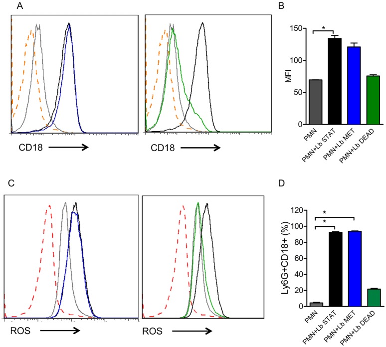
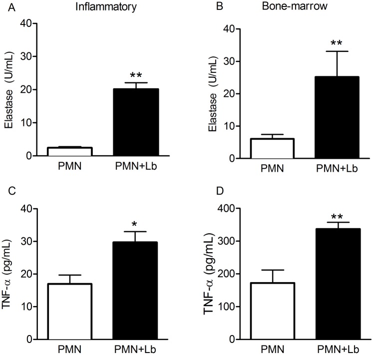
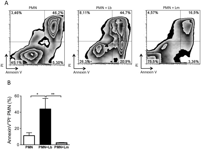
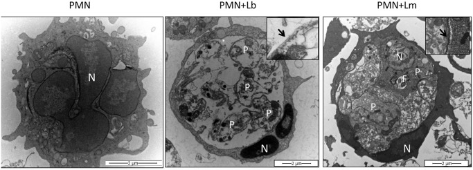
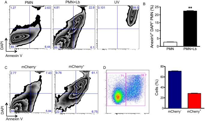
Methods and findings: Inoculation of serum-opsonized L. braziliensis promastigotes in mice induced neutrophil accumulation in vivo, peaking at 24 h. In vitro, exposure of thyoglycollate-elicited inflammatory or bone marrow neutrophils to L. braziliensis modulated the expression of surface molecules such as CD18 and CD62L, and induced the oxidative burst. Using mCherry-expressing L. braziliensis, we determined that such effects were mainly observed in infected and not in bystander cells. Neutrophil activation following contact with L. braziliensis was also confirmed by the release of TNF-α and neutrophil elastase. Lastly, neutrophils infected with L. braziliensis but not with L. major displayed markers of early apoptosis.

Conclusions: We show that L. braziliensis induces neutrophil recruitment in vivo and that neutrophils exposed to the parasite in vitro respond through activation and release of inflammatory mediators. This outcome may impact on parasite elimination, particularly at the early stages of infection.


Conflict of interest statement

The authors have declared that no competing interests exist.

Figures

Fig 1
Fig 1. Neutrophil recruitment following L. braziliensis inoculation.
Individual BALB/c mice were injected with L. braziliensis in the ear dermis. At different time points, cells were prepared and stained for Ly6G and analyzed by flow cytometry. (A) Representative dot plots and gating strategy of neutrophils. (B) Bar graph representation of the number of Ly6G+ cells in naïve mice (grey bars), in mice inoculated with saline (white bars) or inoculated with L. braziliensis (black bars). Data shown (mean ± SEM) are from one experiment representative of two. ***p <0.001, *p<0.05.
Fig 2
Fig 2. Inflammatory neutrophils infected with L. braziliensis up-regulate CD18 and down-regulate CD62L.
Inflammatory neutrophils were co-cultured with mCherry L. braziliensis. (A) Representative dot plot and bar graph depicting the percentage of non-infected (mCherry-) and infected (mCherry+) neutrophils. Representative histograms depicting CD18 (B) and CD62L (C) staining in control neutrophils cultured alone (gray), bystander neutrophils (mCherry-) (blue, shown as% of Max) and infected neutrophils (mCherry+) (red, shown as% of Max). Bar graphs represent the percentages of control, bystander and infected neutrophils expressing CD18 (B) and CD62L (C). Data shown (mean ± SEM) are pooled from two independent repeats. **p<0.01; *p<0.05.
Fig 3
Fig 3. Bone-marrow neutrophils infected with L. braziliensis up-regulate CD18 and down-regulate CD62L.
Bone-marrow neutrophils were co-cultured with mCherry L. braziliensis. (A) Representative dot plot and bar graph representing the percentage of non-infected (mCherry-) and infected (mCherry+) neutrophils. Representative histograms depicting CD18 (B) and CD62L (C) staining in control neutrophils cultured alone (gray), bystander neutrophils (mCherry-) (blue, shown as% of Max) and infected neutrophils (mCherry+) (red, shown as% of Max). Bar graphs represent the percentages of control, bystander and infected neutrophils expressing CD18 (B) and CD62L (C). Data shown (mean ± SEM) are pooled from two independent repeats. *p<0.05.
Fig 4
Fig 4. Exposure to L. braziliensis induces ROS production.
Neutrophils were co-cultured with mCherry L. braziliensis, cells were stained with DHE and ROS production was analyzed by FACS. (A) Histograms represent inflammatory neutrophils cultured alone (gray), bystander neutrophils (mCherry-) (blue) and infected neutrophils (mCherry+) (red). (B) Histograms represent bone marrow neutrophils cultured alone (gray), bystander neutrophils (mCherry-) (blue) and infected neutrophils (mCherry+). Bar graphs represent the MFI of neutrophils cultured alone (PMN), bystanders (mCherry-) (blue) or L. braziliensis-infected (mCherry+) (red). Data (mean ± SEM) are from one experiment representative of two. *p<0.05.
Fig 5
Fig 5. Exposure to dead L. braziliensis does not modulate CD18 expression or ROS production.
Inflammatory neutrophils were co-cultured with L. braziliensis and stained with anti-CD62L. (A,C) Representative histograms depicting ROS production or CD18 staining in neutrophils cultured alone (gray), neutrophils exposed to stationary L. braziliensis (black), metacyclic L. braziliensis (blue) or dead L. braziliensis (green). Dotted orange line depicts unstained neutrophils. (B, D). Bar graphs represent the percentage of neutrophils positive for CD18. Data shown (mean ± SEM) are from one experiment representative of two. *p<0.05.
Fig 6
Fig 6. Production of Elastase and TNF-a by L. braziliensis-exposed neutrophils.
Inflammatory or bone marrow neutrophils were co-cultured with L. braziliensis for 24h. Culture supernatants were assayed for the presence of TNF-α (A,B) and for the presence of free elastase activity (C,D). Data shown (mean ± SEM) are pooled from two independent repeats. **p <0.01, *p<0.05.
Fig 7
Fig 7. Exposure to L. braziliensis but not to L. major triggers neutrophils apoptosis.
Inflammatory neutrophils were co-cultured with L. braziliensis. After 18h, neutrophils were labeled with Annexin V and PI and analyzed by FACS. (A) Representative zebra plots of Annexin V+/PI+ neutrophils (PMN) and of neutrophils co-cultured with L. braziliensis (PMN+Lb) or with L. major (PMN+Lm). (B) Percentage of Annexin V+/PI- neutrophils alone (PMN) or neutrophils cultured with parasites (PMN+Lb and PMN+Lm). Data shown (mean ± SEM) are pooled from two independent repeats. *p<0.05; **p <0.01.
Fig 8
Fig 8. Ultrastructural analysis of neutrophils infected with L. braziliensis or L. major.
Inflammatory neutrophils were co-cultured with L. braziliensis (at a 5:1 parasite:cell ratio), for 18 h. Cells were fixed with 2% glutaraldehyde in 0.1 M cacodylate buffer, pH 7.4, and post-fixed in 1% OsO4 and 0.8% potassium ferricyanide and 5 mM calcium chloride in the same buffer. Cells were dehydrated in a graded series of acetone and embedded in Poly/Bed 812 (Polysciences, Inc.) resin. Ultrathin sections were stained with uranyl acetante and lead citrate and examined on a Zeiss109 transmission electron microscope operating at 80 KV. (A) Uninfected neutrophils exhibiting multilobular nucleus (N). (B) Neutrophil infected with L. braziliensis showing condensed nucleus (N) and unpreserved intracellular parasite structures (P). Insert shows Leishmania microtubule (arrow). (C) Neutrophil infected with L. major showing preserved parasite structures (P) and nucleus (N). Insert shows Leishmania microtubule (arrow).
Fig 9
Fig 9. Apoptosis in neutrophils infected with L. braziliensis.
Bone-marrow neutrophils were co-cultured with L. braziliensis. After 18h, neutrophils were labeled with Annexin V and DAPI and analyzed by FACS. (A) Representative zebra plots of neutrophils (PMN), neutrophils cultured with L. braziliensis (PMN+Lb) or neutrophils exposed to UV (UV). (B) Percentage of DAPI+/Annexin V+ neutrophils (PMN) or neutrophils cultivated with L. braziliensis (PMN+Lb). (C) Representative zebra plots of bystander neutrophils (mCherry-) and of infected neutrophils (mCherry+) expressing DAPI and Annexin V. (D) Representative dot plots of neutrophils infected with mCherry L. braziliensis after 18h, bar graph represents the percentage of non-infected (mCherry-) and infected (mCherry+) neutrophils. Data (mean ± SEM) are from one experiment representative of three experiments. **p <0.01.

Similar articles

Cited by

References

    1. Nathan C (2006) Neutrophils and immunity: challenges and opportunities. Nat Rev Immunol 6: 173–182. - PubMed
    1. Zarbock A, Ley K (2008) Mechanisms and consequences of neutrophil interaction with the endothelium. Am J Pathol 172: 1–7. - PMC - PubMed
    1. Babior BM (1999) NADPH oxidase: an update. Blood 93: 1464–1476. - PubMed
    1. Borregaard N, Sorensen OE, Theilgaard-Monch K (2007) Neutrophil granules: a library of innate immunity proteins. Trends Immunol 28: 340–345. - PubMed
    1. Savill J, Haslett C (1995) Granulocyte clearance by apoptosis in the resolution of inflammation. Semin Cell Biol 6: 385–393. - PubMed

LinkOut - more resources