. 2015 Mar 10;10(3):e0116667.
doi: 10.1371/journal.pone.0116667. eCollection 2015.

Reduced expression of CD27 by collagenase treatment: implications for interpreting b cell data in tissues

Affiliations

Reduced expression of CD27 by collagenase treatment: implications for interpreting b cell data in tissues

Chanjuan Shen et al. PLoS One. .

Abstract

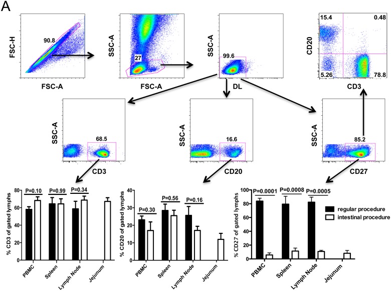
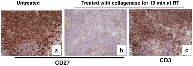
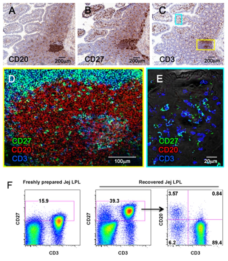
Surface markers have been used to identify distinct cell subpopulations and to delineate various stages of maturation or activation of lymphocytes. In particular CD27 is used for delineation of naïve and memory B cell populations, and is readily detected by flow cytometry. We here used flow cytometry to examine the expression of CD27 on lymphocytes isolated from various tissues of rhesus macaques, and found its expression was consistently low to absent on intestinal cell suspensions. However, immunohistochemistry revealed abundant CD27+ cells in intestinal tissue sections. Further investigation showed the marked loss of CD27 expression on processed intestinal cells was due to collagenase digestion of intestinal tissues, yet CD27 expression was recoverable within hours of cell isolation. By combining confocal microscopy, we confirmed that only a fraction of B cells express CD27, in contrast to expression on all T cells from tissues examined including the gut. Taken together, our results suggest that CD27 may be a memory marker for B cells, but not for T cells, since essentially all CD3 T cells expressed CD27. In summary, it is important to consider the influence of isolation procedures on cell surface expression of phenotypic markers, especially when examining tissue-resident lymphocytes by flow cytometry.


Conflict of interest statement

Competing Interests: The authors have declared no competing interests exist.

Figures

Fig 1
Fig 1. Expression of CD27 on lymphocytes isolated from various tissues of rhesus macaques detected by flow cytometry.
(A) Representative plots generated from whole blood samples showing gating strategy for analysis of CD27+ lymphocytes isolated from all tissues. Specifically, singlets were gated first followed by lymphocytes, and then live lymphocytes (DLneg) were selected for further analysis of CD3+, CD20+, CD27+ as shown as in A; (B) Comparison of percentages of various cells including CD3+, CD20+ and CD27+ after different processing of the same tissues and gating through live lymphocytes. Note that significantly decreased CD27 expression is only detected on lymphocytes subjected to collagenase digestion procedures when compared to no digestion. In contrast, there are no changes in expression of CD3 or CD20 on lymphocytes indicating this is selective for CD27. Bars represent mean percentages ± SEM in each group (n = 7), P values ≤ 0.05 were considered statistically significant.
Fig 2
Fig 2. Comparison of CD27 expression in lymph node tissue sections before and after collagenase treatment.
Note marked loss of CD27 signal is detected following collagenase treatment of adjacent frozen sections of the same lymph node (a and b). In contrast, collagenase treatment has no effect on CD3 expression (c).
Fig 3
Fig 3. CD27 expression recovers on lymphocytes following treatment with collagenase.
(A) Dot plots show collagenase treated PBMCs lose surface CD27 expression, but expression is almost entirely recovered after 4, and entirely recovered after 24 hrs rest / incubation. Numbers in each quadrant indicate the percentage of total live lymphocytes as indicated in Fig. 1. (B) Rapid recovery of CD27 expression on collagenase treated cells occurs in all tissues including intestinal tissues. Histograms are representative of four independent experiments.
Fig 4
Fig 4. Identification and localization of CD27+ cell subsets in intestinal tissues.
Immunohistochemistry staining intestinal B cells (CD20+) (A), CD27+ cells (B), and T cells (CD3+) (C) on the serial adjacent sections. Three-color confocal microscopy images show that CD27+ B cells are mainly located in lymphoid aggregates in the deeper lamina propria of the gut (D), whereas CD27+ cells in the intestinal villi are all T cells (E). Flow cytometry of intestinal cell suspensions confirms most CD27+ cells are CD3+ T cells, with fewer percentages of CD20+ B cells co-expressing CD27 (F).

Similar articles

Cited by

References

    1. Shin H, Iwasaki A (2013) Tissue-resident memory T cells. Immunol Rev 255: 165–181. 10.1111/imr.12087 - DOI - PMC - PubMed
    1. Cauley LS, Lefrancois L (2013) Guarding the perimeter: protection of the mucosa by tissue-resident memory T cells. Mucosal Immunol 6: 14–23. 10.1038/mi.2012.96 - DOI - PMC - PubMed
    1. Hendriks J, Gravestein LA, Tesselaar K, van Lier RA, Schumacher TN, et al. (2000) CD27 is required for generation and long-term maintenance of T cell immunity. Nat Immunol 1: 433–440. - PubMed
    1. Hintzen RQ, Lens SM, Lammers K, Kuiper H, Beckmann MP, et al. (1995) Engagement of CD27 with its ligand CD70 provides a second signal for T cell activation. J Immunol 154: 2612–2623. - PubMed
    1. Nagumo H, Agematsu K, Shinozaki K, Hokibara S, Ito S, et al. (1998) CD27/CD70 interaction augments IgE secretion by promoting the differentiation of memory B cells into plasma cells. J Immunol 161: 6496–6502. - PubMed

Publication types

Substances