. 2015 Mar 10;10(3):e0119727.
doi: 10.1371/journal.pone.0119727. eCollection 2015.

Innate immune response to Streptococcus pyogenes depends on the combined activation of TLR13 and TLR2

Affiliations

Innate immune response to Streptococcus pyogenes depends on the combined activation of TLR13 and TLR2

Christina Fieber et al. PLoS One. .

Abstract

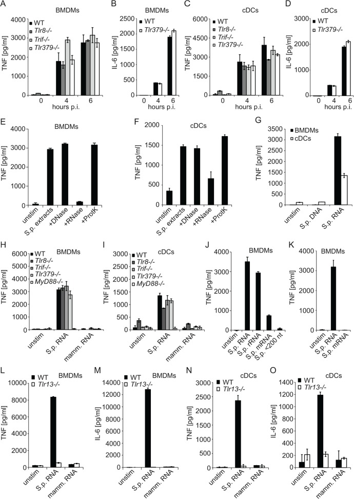
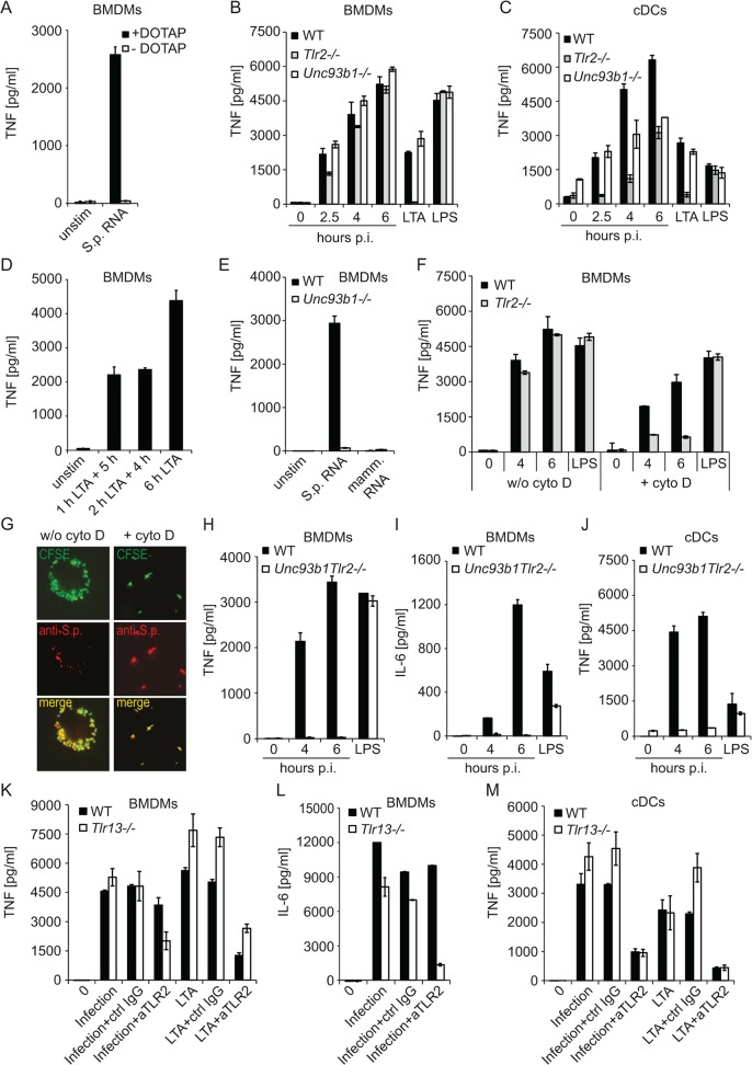
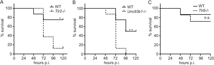
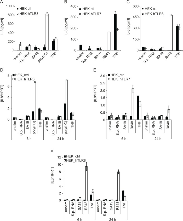
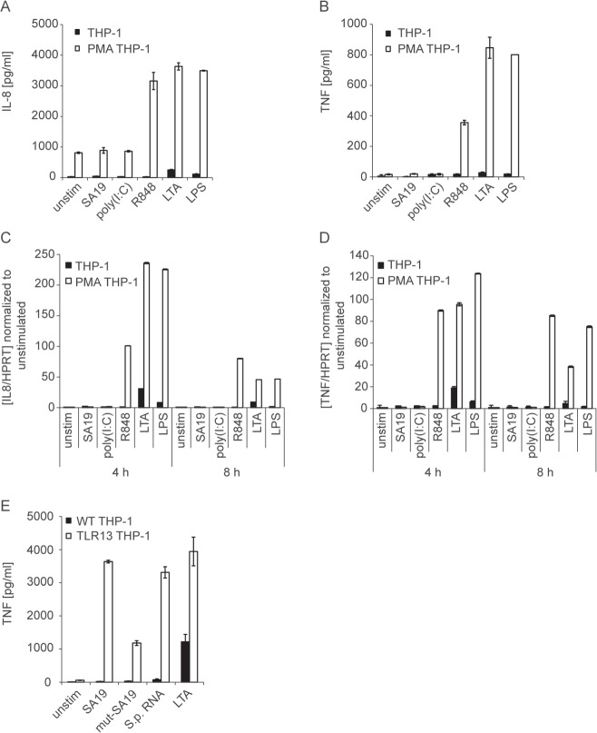
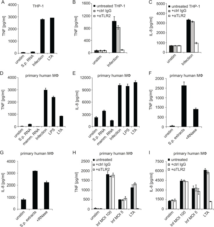
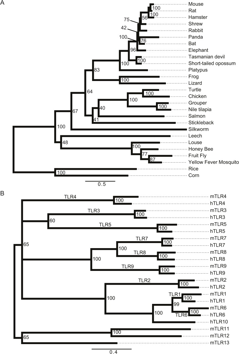
Innate immune recognition of the major human-specific Gram-positive pathogen Streptococcus pyogenes is not understood. Here we show that mice employ Toll-like receptor (TLR) 2- and TLR13-mediated recognition of S. pyogenes. These TLR pathways are non-redundant in the in vivo context of animal infection, but are largely redundant in vitro, as only inactivation of both of them abolishes inflammatory cytokine production by macrophages and dendritic cells infected with S. pyogenes. Mechanistically, S. pyogenes is initially recognized in a phagocytosis-independent manner by TLR2 and subsequently by TLR13 upon internalization. We show that the TLR13 response is specifically triggered by S. pyogenes rRNA and that Tlr13-/- cells respond to S. pyogenes infection solely by engagement of TLR2. TLR13 is absent from humans and, remarkably, we find no equivalent route for S. pyogenes RNA recognition in human macrophages. Phylogenetic analysis reveals that TLR13 occurs in all kingdoms but only in few mammals, including mice and rats, which are naturally resistant against S. pyogenes. Our study establishes that the dissimilar expression of TLR13 in mice and humans has functional consequences for recognition of S. pyogenes in these organisms.


Conflict of interest statement

Competing Interests: The authors declare no financial or commercial conflict of interest.

Figures

Fig 1
Fig 1. S. pyogenes rRNA induces cytokine production in a TLR13-dependent way and independently of TRIF, TLR3, TLR7, TLR8 and TLR9.
BMDMs (A, B) or cDCs (C, D) from Tlr8−/−, Trif−/− or Tlr379 triple-deficient mice as well as control mice (WT) were infected with S. pyogenes (MOI = 50) or left uninfected. At indicated time points, supernatants were collected and TNF (A, C) or IL-6 (B, D) release was measured by ELISA. (E, F) S. pyogenes cells were sonicated and the extracts were treated with either DNase I, RNase A, proteinase K, or left untreated (control extract). These extracts were delivered into BMDMs (E) or cDCs (F) using DOTAP. After stimulation for 6 h, supernatants were collected and TNF release was measured by ELISA. (G) RNA or DNA isolated from S. pyogenes was transfected into WT BMDMs or cDCs and after 6 h of stimulation supernatants were collected and TNF release was measured by ELISA. (H, I) BMDMs (H) and cDCs (I) from Tlr8−/−, Trif−/−, MyD88−/− and Tlr379 triple-deficient mice as well as control mice (WT) were transfected with S. pyogenes or mammalian RNA or left untreated. Supernatants were collected after 6 h and TNF release was measured by ELISA. (J) S. pyogenes total RNA as well as the mRNA (isolated with the MICROBExpress Bacterial mRNA Enrichment Kit, contains 0.3% rRNA), rRNA and nt>200 fractions were used for transfection of BMDMs. TNF release was determined in supernatants of cells after 6 h of stimulation using ELISA. (K) S. pyogenes total RNA as well as the mRNA (isolated with the Ribo-Zero rRNA Removal Kit) were transfected into BMDMs. TNF release was measured 6 h after stimulation using ELISA. (L—O) BMDMs (L, M) and cDCs (N, O) from Tlr13−/− mice as well as control mice (WT) were transfected with S. pyogenes or mammalian RNA or left untreated. Supernatants were collected after 6 h and TNF (L, N) or IL-6 (M, O) release was measured by ELISA. Error bars in all panels represent SDs (n≥3).
Fig 2
Fig 2. S. pyogenes is recognized by a combination of Tlr2- and Tlr13-mediated sensing.
(A) S. pyogenes RNA was either added directly to the BMDMs or transfected into BMDMs using DOTAP. Supernatants were collected after 6 h and TNF release was measured by ELISA. (B, C) BMDMs (B) or cDCs (C) from Tlr2−/− and Unc93b1−/− mice as well as control mice (WT) were infected with S. pyogenes (MOI = 50) or left uninfected, or treated with LTA or LPS as a control. At indicated time points, supernatants were collected and TNF release was measured by ELISA. (D) BMDMs were stimulated for 1 or 2 h with LTA, and after medium change cells were incubated without LTA for additional 5 or 4 h, respectively, As a control, BMDMs were treated with LTA for 6 h. TNF was determined in the supernatants by ELISA. (E) BMDMs from Unc93b1−/− mice as well as control mice (WT) were transfected with streptococcal- or mammalian RNA. Supernatants were collected after 6 h and TNF release was measured by ELISA. (F) BMDMs from Tlr2−/− as well as control mice (WT) were pre-treated for 30 min with cytochalasin D or left untreated prior infection with S. pyogenes (MOI = 50). At indicated time points, supernatants were collected and TNF release was measured by ELISA. (G) BMDMs were pre-treated with cytochalasin D (right panels) or left untreated (left panels) before infection with CFSE-labeled (green) S. pyogenes (MOI = 50). After 4 h cells were fixed and stained with anti S. pyogenes antibody (anti-S.p.) for extracellular bacteria (red). Immunofluorescence image depicts CFSE labeled bacteria (both extra- and intracellular) in green, extracellular antibody-stained bacteria in red, and a merge of the two channels in showing extracellular bacteria in yellow and intracellular bacteria in green. (H—J) BMDMs (H, I) and cDCs (J) from Unc93b1Tlr2 double-deficient mice as well as control mice (WT) were infected with S. pyogenes (MOI = 50) or left uninfected. At indicated time points, supernatants were collected and TNF (H, J) or IL-6 (I) release was measured by ELISA. LPS treatment (6 h) served as specificity control. (K—M) BMDMs (K, L) and cDCs (M) from Tlr13−/− as well as control mice (WT) were incubated for 30–45 min with anti-TLR2, control IgG or left untreated prior to infection with S. pyogenes (MOI = 100) or stimulation with LTA. Supernatants were collected 4 h after infection and TNF (K, M) or IL-6 (L) release was measured by ELISA. Error bars in all panels represent SDs (n≥3).
Fig 3
Fig 3. Entire activation of the immune responses through both Tlr2- and Unc93b1-dependent pathways is required for protective defense against S. pyogenes in mice.
(AC) Kaplan-Meier survival curves of C57BL/6 and Tlr2−/−, Unc93b1−/− (8 mice per genotype) or Tlr9−/− mice (14 mice per genotype) after subcutaneous infection with 1×10⁸ CFU of S. pyogenes ISS3348. Survival was monitored for 6 days. Significance: * = p<0.05; ** = p<0.01.
Fig 4
Fig 4. Human RNA-recognizing TLRs are not capable of sensing S. pyogenes RNA.
(A, D) HEK293 stably expressing human TLR3 (HEK293-hTLR3) or control HEK293 cells (HEK ctl) were transfected with the TLR3 ligand poly(I:C) (5 μg), the Tlr13 ligand oligoribonucleotide SA19 (5 μg), S. pyogenes RNA (5 μg) using DOTAP or stimulated with human TNF (10 ng/ml). IL-8 release was measured in supernatants 24 h post stimulation by ELISA (A) or the levels of IL8 mRNA were determined by qRT-PCR (normalized to HPRT) (D). Mean values ± SD are shown (n ≥ 3). (B, C, E, F) HEK293XL cells stably expressing human TLR7 (B, E) or TLR8 (C, F) or control HEK293XL cells were transfected with SA19 (5 μg), S. pyogenes RNA (5 μg) or stimulated with the TLR7 and TLR8 ligand R848 (5 μg/ml) or human TNF (10 ng/ml). IL-8 release was measured in supernatants 24 h post stimulation by ELISA (B, C) or the levels of IL8 mRNA were determined by qRT-PCR (normalized to HPRT) (E, F). Mean values ± SD are shown (n ≥ 3).
Fig 5
Fig 5. Macrophage-like differentiated THP-1 cells respond to endosomal TLR ligands but not to the canonical TLR13 ligand.
(A, B) THP-1 cells were either treated with 10 nM PMA or left untreated (as described in Material and Methods) before stimulation with R848 (5 μg/ml), LTA (5 μg/ml), LPS (10 ng/ml) or DOTAP-mediated delivery of poly(I:C) (5 μg). Supernatants were collected after 8 h and IL-8 (A) or TNF (B) release was determined by ELISA. Error bars represent SDs (n≥3). (C, D) RNA was isolated from differentiated or undifferentiated THP-1 cells treated as described in (A) and (B) for 4 or 8 h. IL8 (C) and TNF (D) mRNA levels were determined by qRT-PCR (normalized to HPRT). Mean values ± SD are shown (n ≥ 3). (E) THP-1 cells stably expressing TLR13 (TLR13 THP-1) were stimulated with TLR13 ligand SA19, poor TLR13 ligand (mut-SA19), S. pyogenes RNA or LTA for 4 h. TNF was determined in supernatants by ELISA. Error bars in represent SDs (n≥3).
Fig 6
Fig 6. Human macrophage-like cells respond to S. pyogenes infection by TLR2-dependent signaling but fail to recognize S. pyogenes RNA.
(A) PMA-differentiated THP-1 macrophages were stimulated with S. pyogenes RNA (5 μg, using DOTAP), LTA (5 μg/ml), infected with S. pyogenes (MOI = 100) or left untreated. Supernatants were collected 6 h after treatment and TNF release was measured by ELSA. (B, C) PMA-differentiated THP-1 were incubated with an anti-TLR2 (αTLR2) or control IgG antibody for 30 min or left untreated prior to infection with S. pyogenes (MOI = 100), supernatants were collected 6 h post infection and TNF (B) or IL-8 (C) release was measured by ELISA. (D, E) Primary human macrophages were either transfected with 5 μg S. pyogenes or mammalian RNA using DOTAP, infected with S. pyogenes (MOI = 100), stimulated with LTA (5 μg/ml) or LPS (10 ng/ml) or left untreated. Supernatants were collected after 24 h and TNF (D) or IL-8 (E) release was measured by using ELSA. (F, G) Primary human macrophages were stimulated with S. pyogenes extracts, S. pyogenes extracts treated with RNase (+RNase), or left unstimulated. Supernatants were collected after 6 h and TNF (F) or IL-8 (G) release was measured by ELSA. (H, I) Primary human macrophages were incubated with an anti-TLR2 (αTLR2) or control IgG antibody for 30 min or left untreated prior to infection with S. pyogenes (MOI = 100 and MOI = 5) or stimulation with LTS. Supernatants were collected 6 h post infection and TNF (H) or IL-8 (I) release was measured by ELISA. Error bars in all panels represent SDs (n≥3).
Fig 7
Fig 7. Phylogenetic analysis of TLR13 in species and of the TLR repertoires in humans and mice.
(A) The phylogenetic tree displays a choice of species where we identified an ortholog to TLR13 using the OMA browser and NCBI Blast search. The hits were confirmed with FACT to test whether the predicted orthologs have similar feature architecture as the query protein. Thus, the orthologs are firstly similar in sequence and secondly similar in their feature architecture. Numbers at the branch points indicate bootstrap values. (B) The phylogenetic tree displays evolutionary relationship of human and mouse TLR proteins. The tree was constructed as described in (A). Note that if a particular TLR is found in both species then both orthologs exhibit very close evolutionary relationship (bootstrap value 100). The nucleic acid-sensing TLRs do not cluster in one branch: TLR7, TLR8 and TLR9 are found in a different branch than TLR3, and the mouse-specific TLR13 clusters yet in another branch together non-nucleic acid sensors TLR11 and TLR12. Numbers at the branch points indicate bootstrap values (cut-off > 50).

References

    1. Ronald PC, Beutler B. Plant and animal sensors of conserved microbial signatures. Science. 2010; 330: 1061–1064. 10.1126/science.1189468 - DOI - PubMed
    1. Tsatsaronis JA, Walker MJ, Sanderson-Smith ML. Host responses to group a streptococcus: cell death and inflammation. PLoS Pathog. 2014; 10: e1004266 10.1371/journal.ppat.1004266 - DOI - PMC - PubMed
    1. Fieber C, Kovarik P. Responses of innate immune cells to group A Streptococcus. Front Cell Infect Microbiol. 2014; 4: 140 10.3389/fcimb.2014.00140 - DOI - PMC - PubMed
    1. Johansson L, Thulin P, Low DE, Norrby-Teglund A. Getting under the skin: the immunopathogenesis of Streptococcus pyogenes deep tissue infections. Clin Infect Dis. 2010; 51: 58–65. 10.1086/653116 - DOI - PubMed
    1. Wessels MR. Clinical practice. Streptococcal pharyngitis. N Engl J Med. 2011; 364: 648–655. 10.1056/NEJMcp1009126 - DOI - PubMed

Publication types