. 2015 Mar 10;10(3):e0118906.
doi: 10.1371/journal.pone.0118906. eCollection 2015.

Changes in protein expression in two cholangiocarcinoma cell lines undergoing formation of multicellular tumor spheroids in vitro

Affiliations

Changes in protein expression in two cholangiocarcinoma cell lines undergoing formation of multicellular tumor spheroids in vitro

Carlo Mischiati et al. PLoS One. .

Abstract

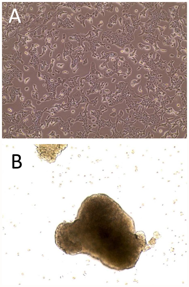
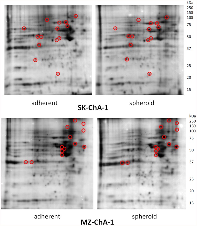
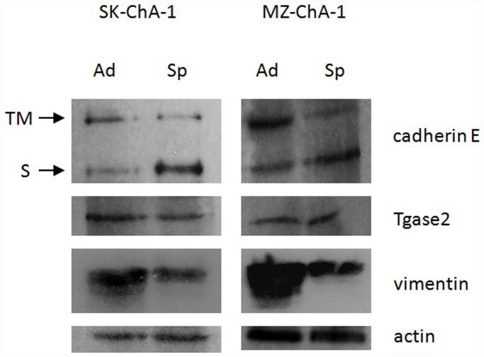
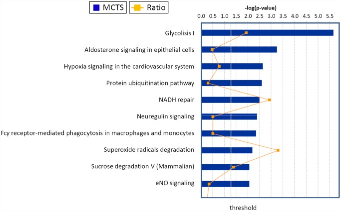
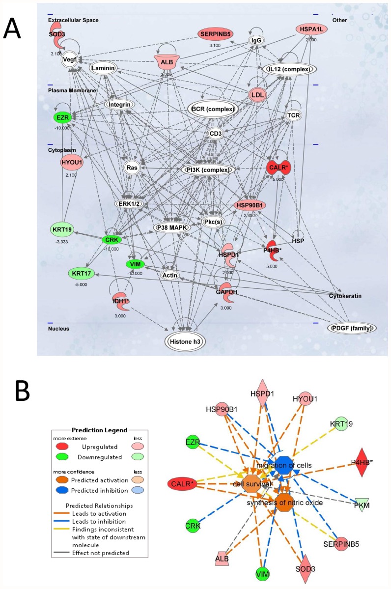
Epithelial-to-Mesenchymal Transition (EMT) is relevant in malignant growth and frequently correlates with worsening disease progression due to its implications in metastases and resistance to therapeutic interventions. Although EMT is known to occur in several types of solid tumors, the information concerning tumors arising from the epithelia of the bile tract is still limited. In order to approach the problem of EMT in cholangiocarcinoma, we decided to investigate the changes in protein expression occurring in two cell lines under conditions leading to growth as adherent monolayers or to formation of multicellular tumor spheroids (MCTS), which are considered culture models that better mimic the growth characteristics of in-vivo solid tumors. In our system, changes in phenotypes occur with only a decrease in transmembrane E-cadherin and vimentin expression, minor changes in the transglutaminase protein/activity but with significant differences in the proteome profiles, with declining and increasing expression in 6 and in 16 proteins identified by mass spectrometry. The arising protein patterns were analyzed based on canonical pathways and network analysis. These results suggest that significant metabolic rearrangements occur during the conversion of cholangiocarcinomas cells to the MCTS phenotype, which most likely affect the carbohydrate metabolism, protein folding, cytoskeletal activity, and tissue sensitivity to oxygen.


Conflict of interest statement

Competing Interests: The commercial funder San Marco Bioenergie did not provide issues of competing interest of any sort (financial, non-financial, professional or personal, including employment, consultancy, patents, products in development, marketed products, etc) nor did it raise restrictions for the authors’ adherence to all PLOS ONE policies on sharing data and materials.

Figures

Fig 1
Fig 1. Morphology of cultures of the SK-ChA-1 cell line.
Panel A, appearance of the adherent bidimensional culture and, Panel B, appearance of the MCTS (Multicellular Tumor Spheroids). In both instances cells were examined by phase contrast microscopy at 10x magnification.
Fig 2
Fig 2. 2D-PAGE separation of proteins contained in the cell lysates of SK-ChA-1 and MZ-ChA-1.
Proteins were first separated by IEF (pH range 3–10) on a non-linear IEF strip (basic pH at the right) followed by SDS-PAGE in the vertical dimension on a 12.5% gel. Circled protein spots were analyzed by mass spectrometry to compare the relative abundance and results were reported in Table 1.
Fig 3
Fig 3. Western blot analysis in adherent and spheroid cells.
The figure presents a representative experiment obtained with SK-ChA-1. Ad: adherent; Sp: spheroid; TM: trans-membrane form of cadherin E; S: soluble form of cadherin E.
Fig 4
Fig 4. Top canonical pathways identified in cholangiocarcinoma cells EMT.
The graph represents host cell pathways with highest score (y-axis) based on the number of differentially regulated proteins using Ingenuity protein analysis. The bar graphs are the pathways most associated with the proteins altered. The orange line graph shows the ratio of the number of molecules from the differentially expressed proteins in EMT that are in the pathway relative to the total number of molecules in the pathway.
Fig 5
Fig 5. Top network functions affected in the EMT process of cholangiocarcinoma cells.
Two relevant networks were generated from EMT-modulated proteins according to the Ingenuity Pathway Knowledge Criteria. A. Cellular compromise, cellular function and maintenance (score = 45). Red, upregulated proteins; green, significantly downregulated proteins; white, proteins known to be in the network but not identified in this study. The color depth indicates the magnitude of the change in protein expression levels. The shape is indicative of the molecular class (i.e protein family). Lines connecting the molecules indicate molecular relationships. Dashed lines indicate indirect interactions, and solid lines indicate direct interactions. The arrow styles indicate specific molecular relationships and the directionality of the interaction. B. Network build up from the three most significant bio-functions (activation Z score >2 or <2): cell survival, synthesis of nitric oxide and migration of cells. The symbols nomenclature is shown in the lower panel.

Similar articles

Cited by

References

    1. Boyer B, Tucker GC, Vallés AM, Franke WW, Thiery JP. Rearrangements of desmosomal and cytoskeletal proteins during the transition from epithelial to fibroblastoid organization in cultured rat bladder carcinoma cells. J Cell Biol. 1989; 109: 1495–1509. - PMC - PubMed
    1. Kalluri R, Weinberg RA. The basics of epithelial-mesenchymal transition. J Clin Invest. 2009; 119: 1420–1428. 10.1172/JCI39104 - DOI - PMC - PubMed
    1. Mathias RA, Simpson RJ. Towards understanding epithelial-mesenchymal transition: a proteomics perspective. Biochim Biophys Acta. 2009; 1794: 1325–1331. 10.1016/j.bbapap.2009.05.001 - DOI - PubMed
    1. Gao D, Vahdat LT, Wong S, Chang JC, Mittal V. Microenvironmental regulation of epithelial-mesenchymal transitions in cancer. Cancer Res. 2012; 72: 4883–4889. 10.1158/0008-5472.CAN-12-1223 - DOI - PMC - PubMed
    1. Nijkamp MM, Span PN, Hoogsteen IJ, van der Kogel AJ, Kaanders JH, et al. Expression of E-cadherin and vimentin correlates with metastasis formation in head and neck squamous cell carcinoma patients. Radiother Oncol. 2011; 99: 344–348. 10.1016/j.radonc.2011.05.066 - DOI - PubMed

Publication types

MeSH terms