. 2015 Mar 10:6:6368.
doi: 10.1038/ncomms7368.

Photonic crystals cause active colour change in chameleons

Affiliations

Photonic crystals cause active colour change in chameleons

Jérémie Teyssier et al. Nat Commun. .

Abstract

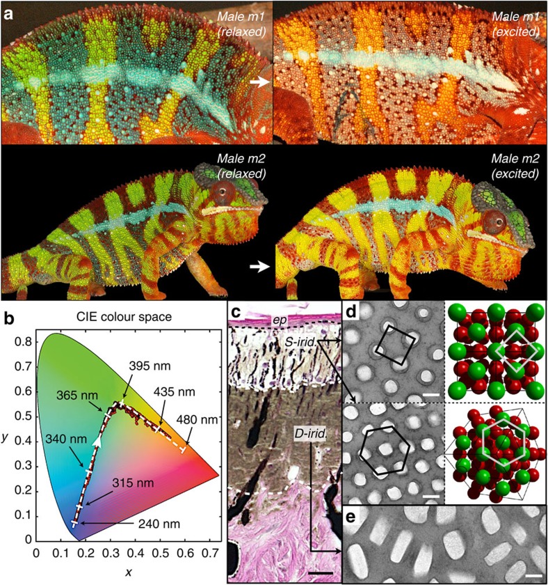
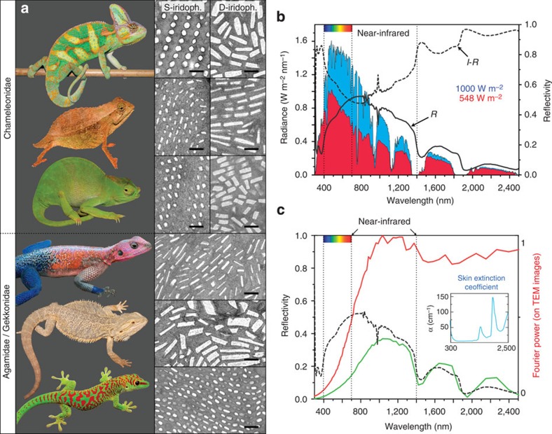
Many chameleons, and panther chameleons in particular, have the remarkable ability to exhibit complex and rapid colour changes during social interactions such as male contests or courtship. It is generally interpreted that these changes are due to dispersion/aggregation of pigment-containing organelles within dermal chromatophores. Here, combining microscopy, photometric videography and photonic band-gap modelling, we show that chameleons shift colour through active tuning of a lattice of guanine nanocrystals within a superficial thick layer of dermal iridophores. In addition, we show that a deeper population of iridophores with larger crystals reflects a substantial proportion of sunlight especially in the near-infrared range. The organization of iridophores into two superposed layers constitutes an evolutionary novelty for chameleons, which allows some species to combine efficient camouflage with spectacular display, while potentially providing passive thermal protection.


Figures

Figure 1
Figure 1. Colour change and iridophore types in panther chameleons.
(a) Reversible colour change is shown for two males (m1 and m2): during excitation (white arrows), background skin shifts from the baseline state (green) to yellow/orange and both vertical bars and horizontal mid-body stripe shift from blue to whitish (m1). Some animals (m2 and Supplementary Movie 2) have their blue vertical bars covered by red pigment cells. (b) Red dots: time evolution in the CIE chromaticity chart of a third male with green skin in a high-resolution video (Supplementary Movie 3); dashed white line: optical response in numerical simulations using a face-centred cubic (FCC) lattice of guanine crystals with lattice parameter indicated with black arrows. (c) Haematoxylin and eosin staining of a cross-section of white skin showing the epidermis (ep) and the two thick layers of iridophores (see also Supplementary Fig. 1). (d) TEM images of guanine nanocrystals in S-iridophores in the excited state and three-dimensional model of an FCC lattice (shown in two orientations). (e) TEM image of guanine nanocrystals in D-iridophores. Scale bars, 20 μm (c); 200 nm (d,e).
Figure 2
Figure 2. In-vivo skin colour change in chameleons is reproduced ex vivo.
(a) TEM images of the lattice of guanine nanocrystals in S-iridophores from the same individual in a relaxed and excited state (two biopsies separated by a distance <1 cm, scale bar, 200 nm). This transformation and corresponding optical response is recapitulated ex vivo by manipulation of white skin osmolarity (from 236 to 1,416 mOsm): (b) reflectivity of a skin sample (for clarity, the 19 reflectivity curves are shifted by 0.02 units along the y axis) and (c) time evolution (in the CIE chromaticity chart) of the colour of a single cell (insets i–vi; Supplementary Movie 4); both exhibit a strong blue shift (red dotted arrow in b) as observed in vivo during behavioural colour change. Dashed white line: optical response in numerical simulations (cf. Fig. 1b) with lattice parameter indicated with dashed arrows. Note that increased osmotic pressure corresponds to behavioural relaxation; hence, the reverse order (white arrowhead in CIE colour chart) of red to green to blue time evolution in comparison with Fig. 1b. (d) Variation of simulated colour photonic response for each vertex of the irreducible first Brillouin zone (colour outside of the Brillouin zone indicates the average among all directions) shown for four lattice parameter values (from Supplementary Movie 5) of the modelled photonic crystal. L-U-K-W-X are standard symmetry points.
Figure 3
Figure 3. Iridophore types in lizards and function of D-iridophores in chameleons.
(a) In addition to F. pardalis (Fig. 1), other chameleonidae (top to bottom: Chamaeleo calyptratus, Rhampholeon spectrum and Kinyongia matschiei) exhibit two superposed layers of (S- and D-) iridophores, whereas agamids (the sister group to chameleons) and gekkonids have a single-type iridophore layer (top to bottom: Agama mwanzae, Pogona vitticeps and Phelsuma grandis). Scale bar, 500 nm. (b) Reflectivity (R) of a panther chameleon white skin sample and solar radiation spectrum (blue curve) at sea level (1,000 W m−2); the product of the solar radiation spectrum and (1−R) yields the amount of sun radiation absorbed by deep tissues (red curve, 548 W m−2). (c) The product of the Fourier power spectrum (red curve, computed from TEM images of D-iridophores) and the extinction coefficient of skin (blue inset) yields a predicted reflectivity distribution (green curve) similar to the measured reflectivity spectrum (black dashed curve) of a panther chameleon red skin sample.

References

    1. Ligon R. A. & McGraw K. J. Chameleons communicate with complex colour changes during contests: different body regions convey different information. Biol. Lett. 9, 20130892 (2013). - PMC - PubMed
    1. Stuart-Fox D. & Moussalli A. Selection for social signalling drives the evolution of chameleon colour change. PLoS Biol. 6, e25 (2008). - PMC - PubMed
    1. Ferguson G. W., Murphy J. B., Ramanamanjato J.-B. & Raselimanana A. P. The Panther Chameleon: Color Variation, Natural History, Conservation, and Captive Management. 118, Krieger Publishing Company (2004).
    1. Stuart-Fox D. & Moussalli A. Camouflage, communication and thermoregulation: lessons from colour changing organisms. Philos. Trans. R. Soc. Lond. B. Biol. Sci. 364, 463–470 (2009). - PMC - PubMed
    1. Messenger J. B. Cephalopod chromatophores: neurobiology and natural history. Biol. Rev. Camb. Philos. Soc. 76, 473–528 (2001). - PubMed

Publication types