. 2015 May;169(4):552-64.
doi: 10.1111/bjh.13335. Epub 2015 Mar 9.

CXCR2 modulates bone marrow vascular repair and haematopoietic recovery post-transplant

Affiliations

CXCR2 modulates bone marrow vascular repair and haematopoietic recovery post-transplant

Sarah J M Hale et al. Br J Haematol. 2015 May.

Abstract

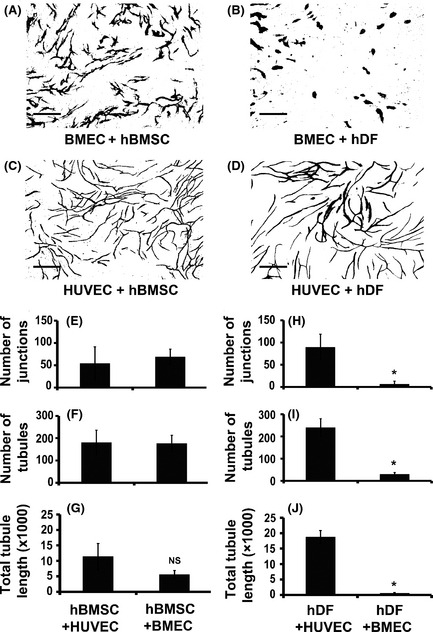
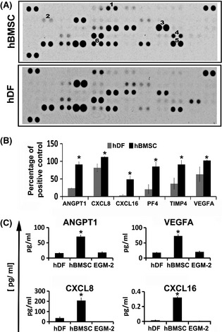
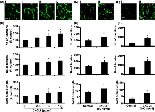
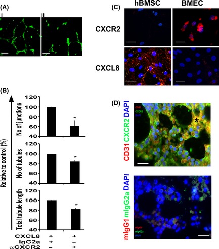
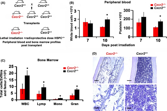
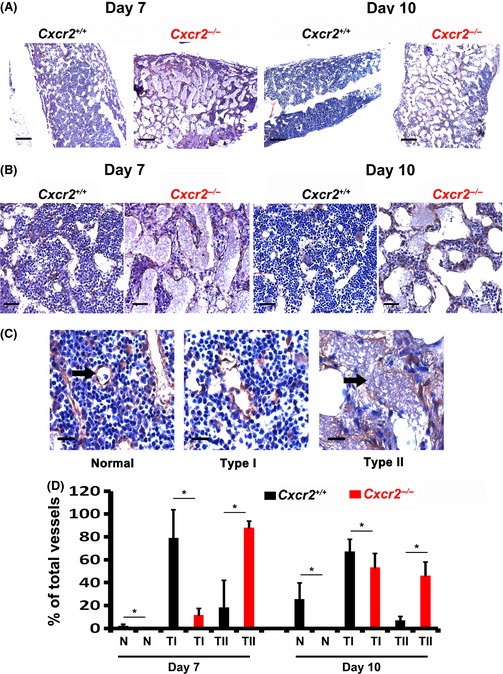
Murine models of bone marrow transplantation show that pre-conditioning regimens affect the integrity of the bone marrow endothelium and that the repair of this vascular niche is an essential pre-requisite for successful haematopoietic stem and progenitor cell engraftment. Little is known about the angiogenic pathways that play a role in the repair of the human bone marrow vascular niche. We therefore established an in vitro humanized model, composed of bone marrow stromal and endothelial cells and have identified several pro-angiogenic factors, VEGFA, ANGPT1, CXCL8 and CXCL16, produced by the stromal component of this niche. We demonstrate for the first time that addition of CXCL8 or inhibition of its receptor, CXCR2, modulates blood vessel formation in our bone marrow endothelial niche model. Compared to wild type, Cxcr2(-/-) mice displayed a reduction in bone marrow cellularity and delayed platelet and leucocyte recovery following myeloablation and bone marrow transplantation. The delay in bone marrow recovery correlated with impaired bone marrow vascular repair. Taken together, our data demonstrate that CXCR2 regulates bone marrow blood vessel repair/regeneration and haematopoietic recovery, and clinically may be a therapeutic target for improving bone marrow transplantation.

Keywords: CXCL8; CXCR2; bone marrow transplantation; bone marrow vascular niche; stem cell niche.


Figures

Figure 1
Figure 1
Human bone marrow stromal cells but not human dermal fibroblasts (hDF) support tubule formation of bone marrow endothelial cells. Human bone marrow stromal cells (hBMSC) or hDF were co‐cultured with either human bone marrow endothelial cell (BMEC) or human umbilical vein endothelial cells (HUVEC) for 14 d in complete Endothelial Cell Growth Medium‐2 supplemented with 8% (v/v) fetal calf serum. Vessel networks were visualized by staining with CD31 antibody. Representative images of vessel formation (scale bars 500 μm) are shown from co‐cultures of BMEC with hBMSC (A), BMEC with hDF (B), HUVEC with hBMSC (C) and HUVEC with hDF (D) at optimal cell ratios (6 × 103 BMEC/HUVEC: 4 × 104 hBMSC and 1·5 × 103 BMEC/HUVEC: 1 × 104 hDF). Quantification of number of junctions (branch points; E), number of tubules (F) and total tubule length (G) for HUVEC or BMEC co‐cultured with hBMSC and number of junctions (H), number of tubules (I) and total tubule length (J) for HUVEC or BMEC co‐cultured with hDF are shown. Results were analysed using Student's t‐test, *P ≤ 0·05. Data are mean ± standard deviation of at least three separate experiments using at least two separate batches of hDF and hBMSC.
Figure 2
Figure 2
Differential expression of pro‐angiogenic proteins in human bone marrow stromal cells and human dermal fibroblasts (hDF). Conditioned media from two separate batches of human bone marrow stromal cells (hBMSC) and hDF were analysed using angiogenic antibody arrays. Representative blots are shown in (A; 1–6 = ANGPT1, CXCL16, CXCL8, PF4, TIMP4 and VEGFA respectively). The mean pixel density was calculated using ImageJ software for each protein tested and the relative percentage of mean pixel density for each protein was calculated after subtraction of the negative control (B). Quantitative ELISAs were performed on the conditioned medium obtained from hBMSC and hDF to compare the levels of ANGPT1, VEGFA, CXCL16 and CXCL8 (C). Results were analysed using Student's t‐test, *P ≤ 0·05. Data are mean ± standard deviation of at least three separate experiments using at least two separate batches of hDF and hBMSC. EGM‐2, Endothelial Cell Growth Medium‐2.
Figure 3
Figure 3
CXCL8 increases vessel formation of human bone marrow endothelial cells. Bone marrow endothelial cells (BMEC) were seeded onto growth factor‐reduced Matrigel and supplemented with increasing doses of cytokine or carrier (0·5% w/v bovine serum albumin) as indicated in the figure. Representative pictures (A: i–iv) of vessel networks formed in Matrigel in response to CXCL8 (scale bars = 200 μmol/l) and (B) quantification of number of junctions, number of tubules, and total tubule length after 16 h incubation are shown. Representative images of vessel formation are also shown from co‐cultures of BMEC with either human bone marrow stromal cells (hBMSC) or human dermal fibroblasts (hDF) incubated in complete EGM‐2 media supplemented with 8% (v/v) fetal calf serum and either carrier or CXCL8 (C: i & ii and E: i & ii respectively; scale bars = 500 μmol/l), Vessel networks were visualized by fluorescence microscopy of eGFP‐labelled BMEC. Quantification of number of junctions (branch points), number of tubules and total tubule length in co‐cultures of BMEC and hBMSC (D) or BMEC and hDF (F) in the presence of carrier or CXCL8 are also shown. Results were analysed using Student's t‐test, *P ≤ 0·05. Data are mean ± standard deviation of at least three separate experiments.
Figure 4
Figure 4
CXCR2 modulates angiogenesis and is expressed on human bone marrow endothelium. eGFP‐labelled human bone marrow endothelial cells (BMEC) were seeded onto growth factor‐reduced Matrigel and supplemented with CXCL8 (5 μg/ml) in the presence of either CXCR2 neutralizing antibody (20 μg/ml) or isotype control (IgG2a, 20 μg/ml). Representative pictures of vessel networks formed after 16 h either in presence of isotype control or anti‐CXCR2 neutralizing antibody are shown (A: i and ii respectively). Quantification of number of junctions (branch points), number of tubules and total tubule length in the absence or presence of CXCR2 neutralizing antibody (αCXCR2) is shown (B). Representative pictures of BMEC or hBMSC stained with either CXCR2 or CXCL8 (C; scale bars = 200 μm) and human normal bone marrow biopsies co‐stained with anti CD31 (red) and CXCR2 (green) antibodies (D; *sinusoid, scale bars = 50 μm). Cell nuclei were stained with DAPI (blue). Results were analysed using Student's t‐test, *P ≤ 0·05. Data are mean ± standard deviation of at least three independent experiments.
Figure 5
Figure 5
Delayed haematopoietic reconstitution in Cxcr2 knockout animals. B6.129S2(C)‐Cxcr2 m1Mwm/J mice, heterozygous for Cxcr2 (Il8rb) knockout were obtained from Jackson Laboratories and bred in house to generate Cxcr2 homozygous knockout and littermate wild type controls. Cxcr2 homozygous knockout (−/−) and wild type (+/+) littermate control animals were treated with 9·5 Gy irradiation dose and transplanted within 24 h with a radio‐protective dose (1 × 106) of total wild type bone marrow cells. Peripheral blood samples were taken from animals at days 7 and 10 and haematology analysis performed on a veterinary blood analyser. After 14 d femurs and tibias were harvested and bone marrow extracted before performing haematology analysis. Schematic representation of study (A), comparative white blood cell and platelet counts in peripheral blood (B), bone marrow haematology analysis at day 14 (WBC = white blood cell count, Lymp = lymphocyte, Mono = monocyte and Gran = granulocyte; C), and representative images of low magnification (top panel; scale bars = 200 μm) and higher magnification (bottom panel; scale bar = 50 μm) bone marrow sections stained with haematoxylin to reveal cellularity (D). Data are mean ± standard deviation. n = 13–16 animals per group. Results were analysed using Student's t‐test, *P ≤ 0·05 comparing cell numbers in Cxcr2 −/− versus wild type (Cxcr2 +/+) animals.
Figure 6
Figure 6
Impaired vascular regeneration in Cxcr2 knockout animals. B6.129S2(C)‐Cxcr2 tm1Mwm/J mice, homozygous knockout (Cxcr2 −/−) or wild type (Cxcr2 +/+) littermates were treated with 9·5 Gy irradiation dose and transplanted within 24 h with a radio‐protective dose (1 × 106) of total bone marrow cells. After 7 or 14 d femurs were harvested, fixed and embedded in paraffin. Bone marrow sections were stained with anti‐mouse VE‐Cadherin antibody to highlight bone marrow endothelium. Representative images of recovering bone marrow endothelium at low magnification (A; scale bars = 200 μm) and higher magnification (B; scale bars = 50 μm) post‐irradiation are shown. Representative images of examples of normal (N), or type I (mild/moderately damaged) or type II (significantly damaged) bone marrow vessels (C; scale bars = 25 μm; black arrow) and quantification of these vessel types post‐recovery are shown (D). Data are mean ± standard deviation. n = 5 animals per group. Results were analysed using analysis of variance (anova) and Student's t‐test, *P ≤ 0·05 comparing vessel numbers in Cxcr2 −/− versus wild type (Cxcr2 +/+) animals.

References

    1. Avecilla, S.T. , Hattori, K. , Heissig, B. , Tejada, R. , Liao, F. , Shido, K. , Jin, D.K. , Dias, S. , Zhang, F. , Hartman, T.E. , Hackett, N.R. , Crystal, R.G. , Witte, L. , Hicklin, D.J. , Bohlen, P. , Eaton, D. , Lyden, D. , de Sauvage, F. & Rafii, S. (2004) Chemokine‐mediated interaction of hematopoietic progenitors with the bone marrow vascular niche is required for thrombopoiesis. Nature Medicine, 10, 64–71. - PubMed
    1. Barker, J.E. (1994) Sl/Sld hematopoietic progenitors are deficient in situ. Experimental Hematology, 22, 174–177. - PubMed
    1. Barker, J.E. (1997) Early transplantation to a normal microenvironment prevents the development of Steel hematopoietic stem cell defects. Experimental Hematology, 25, 542–547. - PubMed
    1. de Barros, A.P. , Takiya, C.M. , Garzoni, L.R. , Leal‐Ferreira, M.L. , Dutra, H.S. , Chiarini, L.B. , Meirelles, M.N. , Borojevic, R. & Rossi, M.I. (2010) Osteoblasts and bone marrow mesenchymal stromal cells control hematopoietic stem cell migration and proliferation in 3D in vitro model. PLoS ONE, 5, e9093. - PMC - PubMed
    1. Bikfalvi, A. & Han, Z.C. (1994) Angiogenic factors are hematopoietic growth factors and vice versa. Leukemia, 8, 523–529. - PubMed

Publication types

Substances