

Sinus of valsalva aneurysms: review of the literature and an update on management
- PMID: 25757442
- PMCID: PMC6711005
- DOI: 10.1002/clc.22359
Sinus of valsalva aneurysms: review of the literature and an update on management
Abstract
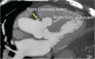
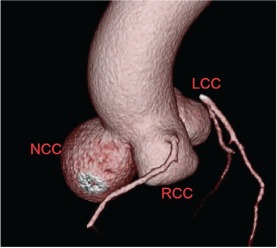
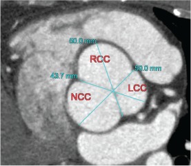
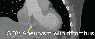
Sinus of Valsalva aneurysm (SOVA), a congenital or acquired cardiac defect that is present in roughly 0.09% of the general population, often presents as an incidental finding during cardiac imaging. Although an echocardiogram is the standard imaging technique for such findings, cardiac computed tomography angiography (CCTA) has been increasingly utilized. If SOVA is diagnosed, CCTA is also a useful test for patients who are at low to intermediate risk for coronary artery disease (CAD) prior to surgical repair. CCTA can accurately rule out CAD, obviating the need for invasive angiography in most cases, which may be more risky in SOVA patients because their coronaries may be more difficult to engage and their aortic root may be more prone to injury. Although surgery has previously been the treatment of choice, transcatheter techniques have added to the spectrum of nonsurgical alternatives for repair. We report here 4 incidental SOVA cases and review the current literature.
© 2015 Wiley Periodicals, Inc.
Figures
References
-
- Bricker AO, Avutu B, Mohammed TL, et al. Valsalva sinus aneurysms: findings at CT and MR imaging. Radiographics. 2010;30:99–110. - PubMed
-
- Troupis JM, Nasis A, Pasricha S, et al. Sinus valsalva aneurysm on cardiac CT angiography: assessment and detection. J Med Imaging Radiat Oncol. 2013;57:444–447. - PubMed
-
- Ott DA. Aneurysm of the sinus of Valsalva. Semin Thorac Cardiovasc Surg Pediatr Card Surg Annu. 2006:165–176. - PubMed
-
- Edwards JE, Burchell HB. Specimen exhibiting the essential lesion in aneurysm of the aortic sinus. Proc Staff Meet Mayo Clin. 1956;31:407–412. - PubMed
-
- Moustafa S, Mookadam F, Cooper L, et al. Sinus of valsalva aneurysms—47 years of a single center experience and systematic overview of published reports. Am J Cardiol. 2007;99:1159–1164. - PubMed
Publication types
MeSH terms
LinkOut - more resources
Full Text Sources
Other Literature Sources
Medical
Miscellaneous
