

Efficient inversions and duplications of mammalian regulatory DNA elements and gene clusters by CRISPR/Cas9
- PMID: 25757625
- PMCID: PMC4524425
- DOI: 10.1093/jmcb/mjv016
Efficient inversions and duplications of mammalian regulatory DNA elements and gene clusters by CRISPR/Cas9
Abstract
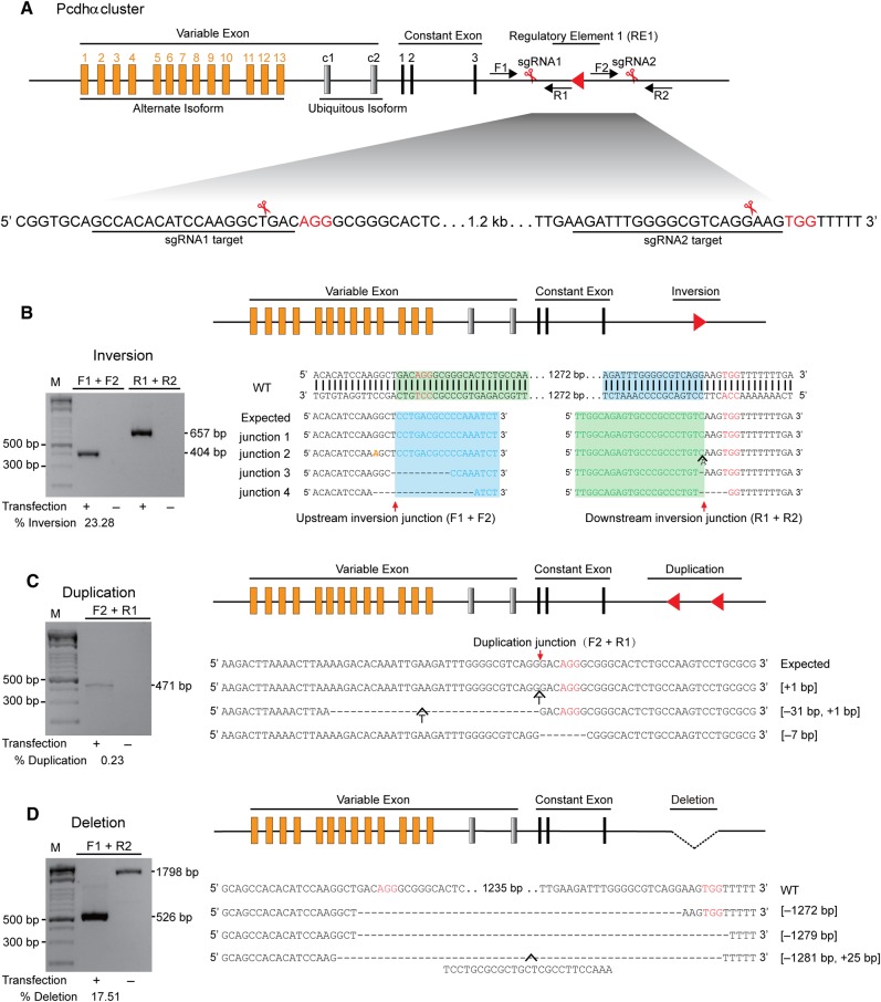
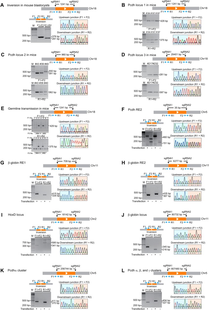
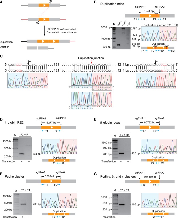
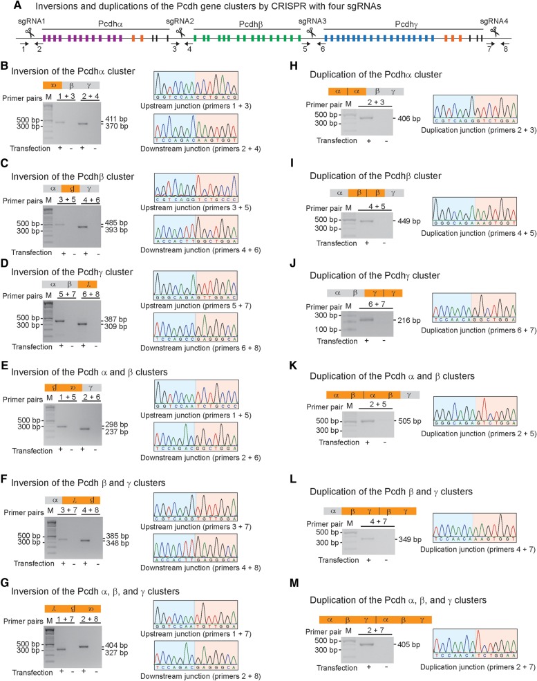
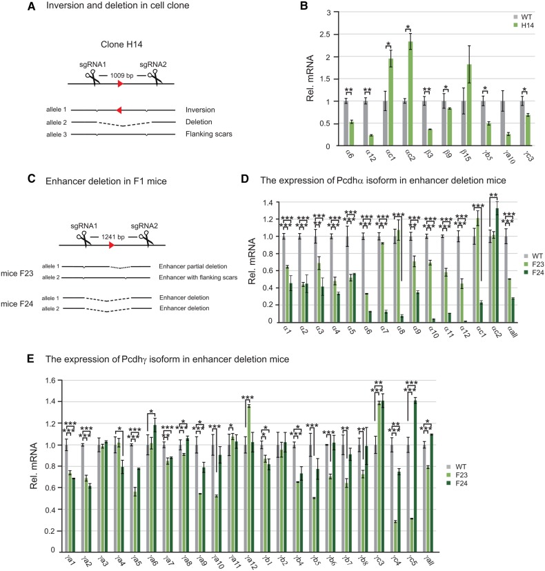
The human genome contains millions of DNA regulatory elements and a large number of gene clusters, most of which have not been tested experimentally. The clustered regularly interspaced short palindromic repeats (CRISPR)/CRISPR-associated nuclease 9 (Cas9) programed with a synthetic single-guide RNA (sgRNA) emerges as a method for genome editing in virtually any organisms. Here we report that targeted DNA fragment inversions and duplications could easily be achieved in human and mouse genomes by CRISPR with two sgRNAs. Specifically, we found that, in cultured human cells and mice, efficient precise inversions of DNA fragments ranging in size from a few tens of bp to hundreds of kb could be generated. In addition, DNA fragment duplications and deletions could also be generated by CRISPR through trans-allelic recombination between the Cas9-induced double-strand breaks (DSBs) on two homologous chromosomes (chromatids). Moreover, junctions of combinatorial inversions and duplications of the protocadherin (Pcdh) gene clusters induced by Cas9 with four sgRNAs could be detected. In mice, we obtained founders with alleles of precise inversions, duplications, and deletions of DNA fragments of variable sizes by CRISPR. Interestingly, we found that very efficient inversions were mediated by microhomology-mediated end joining (MMEJ) through short inverted repeats. We showed for the first time that DNA fragment inversions could be transmitted through germlines in mice. Finally, we applied this CRISPR method to a regulatory element of the Pcdhα cluster and found a new role in the regulation of members of the Pcdhγ cluster. This simple and efficient method should be useful in manipulating mammalian genomes to study millions of regulatory DNA elements as well as vast numbers of gene clusters.
Keywords: CRISPR/Cas9; DNA regulatory element inversion; deletion; duplication; enhancer; gene cluster; genome manipulation.
© The Author (2015). Published by Oxford University Press on behalf of Journal of Molecular Cell Biology, IBCB, SIBS, CAS.
Figures
Similar articles
-
Precise and Predictable CRISPR Chromosomal Rearrangements Reveal Principles of Cas9-Mediated Nucleotide Insertion.Mol Cell. 2018 Aug 16;71(4):498-509.e4. doi: 10.1016/j.molcel.2018.06.021. Epub 2018 Jul 19. Mol Cell. 2018. PMID: 30033371
-
Generation of megabase-scale deletions, inversions and duplications involving the Contactin-6 gene in mice by CRISPR/Cas9 technology.BMC Genet. 2017 Dec 28;18(Suppl 1):112. doi: 10.1186/s12863-017-0582-7. BMC Genet. 2017. PMID: 29297312 Free PMC article.
-
Characterization of genomic deletion efficiency mediated by clustered regularly interspaced short palindromic repeats (CRISPR)/Cas9 nuclease system in mammalian cells.J Biol Chem. 2014 Aug 1;289(31):21312-24. doi: 10.1074/jbc.M114.564625. Epub 2014 Jun 6. J Biol Chem. 2014. PMID: 24907273 Free PMC article.
-
DNA fragment editing of genomes by CRISPR/Cas9.Yi Chuan. 2015 Oct;37(10):992-1002. doi: 10.16288/j.yczz.15-291. Yi Chuan. 2015. PMID: 26496751 Review.
-
CRISPR/Cas9 Technology in Translational Biomedicine.Cell Physiol Biochem. 2020 Apr 17;54(3):354-370. doi: 10.33594/000000224. Cell Physiol Biochem. 2020. PMID: 32298553 Review.
Cited by
-
A CRISPR Path to Engineering New Genetic Mouse Models for Cardiovascular Research.Arterioscler Thromb Vasc Biol. 2016 Jun;36(6):1058-75. doi: 10.1161/ATVBAHA.116.304790. Epub 2016 Apr 21. Arterioscler Thromb Vasc Biol. 2016. PMID: 27102963 Free PMC article. Review.
-
Progress in and Prospects of Genome Editing Tools for Human Disease Model Development and Therapeutic Applications.Genes (Basel). 2023 Feb 14;14(2):483. doi: 10.3390/genes14020483. Genes (Basel). 2023. PMID: 36833410 Free PMC article. Review.
-
CRISPR/Cas9 and FLP-FRT mediated regulatory dissection of the BX-C of Drosophila melanogaster.Chromosome Res. 2023 Jan 31;31(1):7. doi: 10.1007/s10577-023-09716-w. Chromosome Res. 2023. PMID: 36719476
-
GRIBCG: a software for selection of sgRNAs in the design of balancer chromosomes.BMC Bioinformatics. 2019 Mar 12;20(1):122. doi: 10.1186/s12859-019-2712-x. BMC Bioinformatics. 2019. PMID: 30866794 Free PMC article.
-
Highly efficient correction of structural mutations of 450 kb KIT locus in kidney cells of Yorkshire pig by CRISPR/Cas9.BMC Mol Cell Biol. 2019 Apr 3;20(1):4. doi: 10.1186/s12860-019-0184-5. BMC Mol Cell Biol. 2019. PMID: 31041890 Free PMC article.
References
-
- Banerji J., Olson L., Schaffner W. (1983). A lymphocyte-specific cellular enhancer is located downstream of the joining region in immunoglobulin heavy chain genes. Cell 33, 729–740. - PubMed
-
- Barrangou R., Fremaux C., Deveau H., et al. (2007). CRISPR provides acquired resistance against viruses in prokaryotes. Science 315, 1709–1712. - PubMed
-
- Blackwood E.M., Kadonaga J.T. (1998). Going the distance: a current view of enhancer action. Science 281, 60–63. - PubMed
Publication types
MeSH terms
Substances
LinkOut - more resources
Full Text Sources
Other Literature Sources
