. 2015 Feb 8;17(1):16.
doi: 10.1186/s13058-015-0517-z.

Lobular breast cancer: molecular basis, mouse and cellular models

Affiliations

Lobular breast cancer: molecular basis, mouse and cellular models

Matthias Christgen et al. Breast Cancer Res. .

Abstract

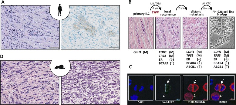
Infiltrating lobular breast cancer (ILC) is the most common special breast cancer subtype. With mutational or epigenetic inactivation of the cell adhesion molecule E-cadherin (CDH1) being confined almost exclusively to ILC, this tumor entity stands out from all other types of breast cancers. The molecular basis of ILC is linked to loss of E-cadherin, as evidenced by human CDH1 germline mutations and conditional knockout mouse models. A better understanding of ILC beyond the level of descriptive studies depends on physiologically relevant and functional tools. This review provides a detailed overview on ILC models, including well-characterized cell lines, xenograft tumors and genetically engineered mouse models. We consider advantages and limitations of these models and evaluate their representativeness for human ILC. The still incompletely defined mechanisms by which loss of E-cadherin drives malignant transformation are discussed based on recent findings in these models. Moreover, candidate genes and signaling pathways potentially involved in ILC development and progression as well as anticancer drug and endocrine resistance are highlighted.


Figures

Figure 1
Figure 1
Infiltrating lobular breast cancer, a human infiltrating lobular breast cancer cell line and a genetically engineered mouse model for infiltrating lobular breast cancer. (A) Representative photomicrographs of infiltrating lobular breast cancer (ILC) stained with hematoxylin and eosin (left) or subjected to immunohistochemistry for E-cadherin (right). Note the E-cadherin-positive normal mammary gland duct surrounded by E-cadherin-negative ILC cells. (B) Molecular evolution of the IPH-926 ILC cell line. Photomicrographs show the histomorphology of the corresponding clinical tumor specimens and the IPH-926 cell line in vitro. Arrow highlights a single file linear cord reminiscent of primary ILC. AI, aromatase inhibitor; CTX, various conventional chemotherapies; LIR, local irradiation; TAM, tamoxifen; TSPP, transition to a secondary pleomorphic phenotype; yrs, years; M, mutational inactivation; ↑, overexpression; ↓, loss of expression. (C) Reconstitution of E-cadherin in ILC cells induces relocation of p120-catenin (p120) to the cell membrane. Shown are fluorescence images of IPH-926 cells transiently transfected with an E-cadherin–enhanced green fluorescent protein (Ecad-EGFP) expression construct and stained with an anti-p120-Alexa647 antibody. Closed arrow, cells with ectopic expression of Ecad-EGFP; open arrow, a cell without Ecad-EGFP. Note the accentuated membranous p120 staining in cells expressing Ecad-EGFP. DAPI, 4′,6-diamidino-2-phenylindole. (D) Mouse ILC from genetically engineered mouse models. Left, a tumor reminiscent of classic ILC; right, a pleomorphic mouse ILC. Both micrographs generated from the WAPcre;Cdh1 F/F ;Trp53 F/F mouse ILC model.

References

    1. Foote FW, Stewart FW. Lobular carcinoma in situ: a rare form of mammary cancer. Am J Pathol. 1941;17:491–6. - PMC - PubMed
    1. Willis RA. Epithelial tumours of the breast. In: Willis RA, editor. Pathology of tumours. London: Butterworth Co. Ltd; 1948. pp. 208–57.
    1. Hyafil F, Babinet C, Jacob F. Cell–cell interactions in early embryogenesis: a molecular approach to the role of calcium. Cell. 1981;26:447–54. doi: 10.1016/0092-8674(81)90214-2. - DOI - PubMed
    1. Niessen CM, Gottardi CJ. Molecular components of the adherens junction. Biochim Biophys Acta. 2008;1778:562–71. doi: 10.1016/j.bbamem.2007.12.015. - DOI - PMC - PubMed
    1. Moll R, Mitze M, Frixen UH, Birchmeier W. Differential loss of E-cadherin expression in infiltrating ductal and lobular breast carcinomas. Am J Pathol. 1993;143:1731–42. - PMC - PubMed

MeSH terms