

Clinical Trial
doi: 10.1038/jcbfm.2015.32.
Epub 2015 Mar 11.
Striatal blood-brain barrier permeability in Parkinson's disease
Affiliations
- PMID: 25757748
- PMCID: PMC4420870
- DOI: 10.1038/jcbfm.2015.32
Item in Clipboard
Clinical Trial
Striatal blood-brain barrier permeability in Parkinson's disease
J Cereb Blood Flow Metab.
2015 May.
Abstract
In vivo studies have shown that blood-brain barrier (BBB) dysfunction is involved in the course of Parkinson's disease (PD). However, these have lacked either anatomic definition or the ability to recognize minute changes in BBB integrity. Here, using histologic markers of serum protein, iron, and erythrocyte extravasation, we have shown significantly increased permeability of the BBB in the postcommissural putamen of PD patients. The dense innervation of the striatum by PD-affected regions allows for exploitation of this permeability for therapeutic goals. These results are also discussed in the context of the retrograde trans-synaptic hypothesis of PD spread.
Figures
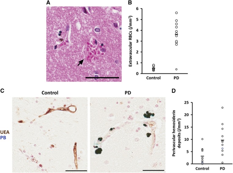
Extravasated erythrocytes and perivascular hemosiderin deposits are more abundant in Parkinson's disease (PD) striatum. (A) Multiple erythrocytes (arrow) lying within the striatal parenchyma adjacent to the wall of a striatal vessel. Scale bar=50 μm. (B) Quantification of extravascular erythrocytes in the striata of control and PD subjects. P<2 × 10⁻⁷. Line represents mean. n=11 controls, 12 PD. (C) Representative micrographs of Prussian Blue staining demonstrating perivascular hemosiderin (dark blue) adjacent to ulex europaeus agglutinin-stained vessels (brown) in PD striatum (right) and lack thereof in control tissue (left). Scale bars=50 μm. (D) Quantification of perivascular hemosiderin in the striata of control and PD subjects. P<0.005. Line represents mean. n=11 controls, 12 PD.
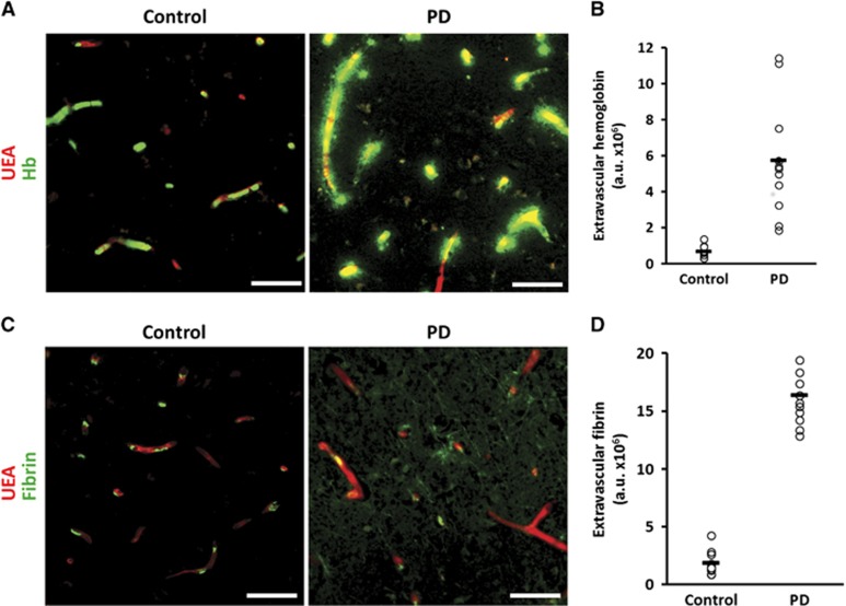
Leakage of serum proteins is increased in Parkinson's disease (PD) striatum. (A) Hemoglobin (Hb; green) and ulex europaeus agglutinin-positive vessel (red) staining in control (left) and PD (right) striatum. Scale bars=50 μm. (B) Quantification of the extravascular portion of Hb staining P<2 × 10⁻⁵. Line represents mean. n=11 controls, 12 PD. (C) Fibrin(ogen) (green) and UEA-positive vessel (red) staining in control (left) and PD (right) striatum. Scale bars=50 μm. (D) Quantification of the extravascular portion of fibrin(ogen) staining P<3 × 10⁻⁵. Line represents mean. n=11 controls, 12 PD.
References
-
- Woulfe JM, Gray MT, Gray DA, Munoz DG, Middeldorp JM. Hypothesis: a role for EBV-induced molecular mimicry in Parkinson's disease. Parkinsonism Relat Disord. 2014;20:685–694. - PubMed
-
- Tanji K, Mori F, Mimura J, Itoh K, Kakita A, Takahashi H, et al. Proteinase K-resistant alpha-synuclein is deposited in presynapses in human Lewy body disease and A53T alpha-synuclein transgenic mice. Acta Neuropathol. 2010;120:145–154. - PubMed
Publication types
MeSH terms
Substances
Grants and funding
LinkOut - more resources
Full Text Sources
Other Literature Sources
Medical
