

Rhabdomyoblastic Differentiation in Head and Neck Malignancies Other Than Rhabdomyosarcoma
- PMID: 25757816
- PMCID: PMC4651923
- DOI: 10.1007/s12105-015-0624-2
Rhabdomyoblastic Differentiation in Head and Neck Malignancies Other Than Rhabdomyosarcoma
Abstract
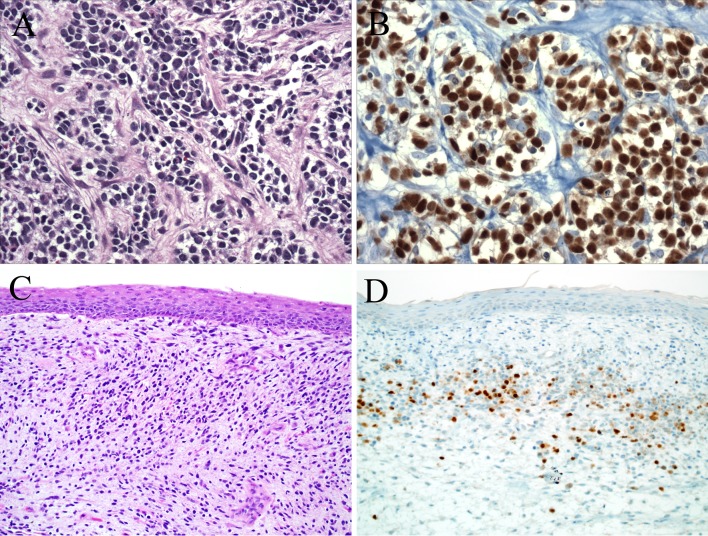
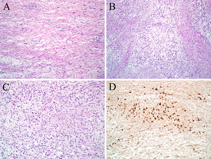
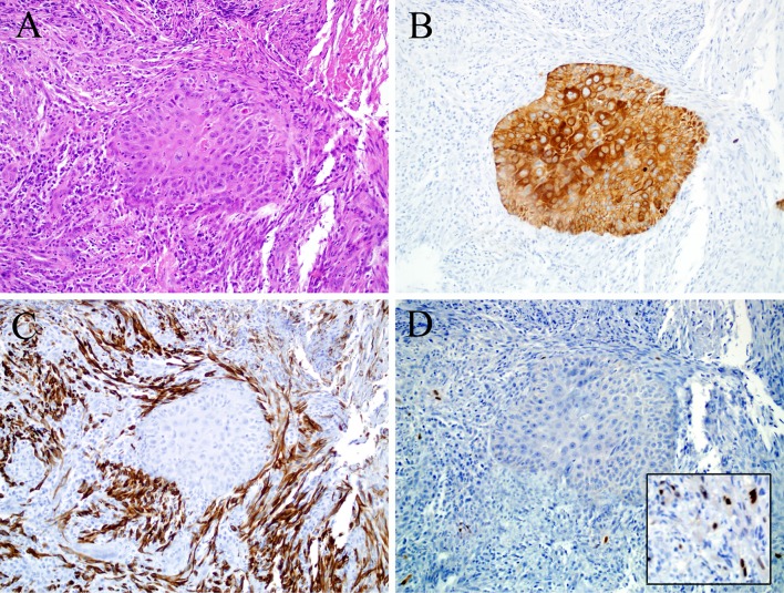
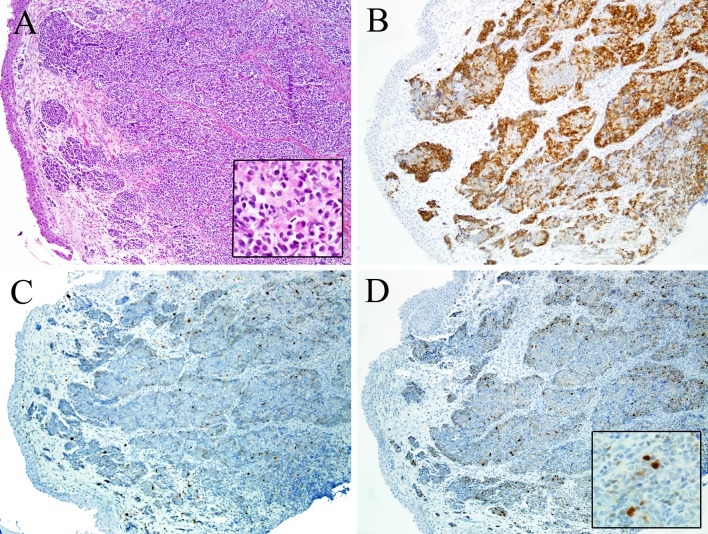
Rhabdomyosarcoma is a relatively common soft tissue sarcoma that frequently affects children and adolescents and may involve the head and neck. Rhabdomyosarcoma is defined by skeletal muscle differentiation which can be suggested by routine histology and confirmed by immunohistochemistry for the skeletal muscle-specific markers myogenin or myoD1. At the same time, it must be remembered that when it comes to head and neck malignancies, skeletal muscle differentiation is not limited to rhabdomyosarcoma. A lack of awareness of this phenomenon could lead to misdiagnosis and, subsequently, inappropriate therapeutic interventions. This review focuses on malignant neoplasms of the head and neck other than rhabdomyosarcoma that may exhibit rhabdomyoblastic differentiation, with an emphasis on strategies to resolve the diagnostic dilemmas these tumors may present. Axiomatically, no primary central nervous system tumors will be discussed.
Keywords: Head and neck malignancies; MyoD1; Myogenin; Rhabdomyoblastic differentiation; Rhabdomyosarcoma; Skeletal muscle differentiation; Soft tissue sarcomas.
Figures
Similar articles
-
Histiocyte-rich rhabdomyoblastic tumor: rhabdomyosarcoma, rhabdomyoma, or rhabdomyoblastic tumor of uncertain malignant potential? A histologically distinctive rhabdomyoblastic tumor in search of a place in the classification of skeletal muscle neoplasms.Mod Pathol. 2019 Mar;32(3):446-457. doi: 10.1038/s41379-018-0145-0. Epub 2018 Oct 4. Mod Pathol. 2019. PMID: 30287926
-
Clinicopathologic features of 300 rhabdomyosarcomas with emphasis upon differential expression of skeletal muscle specific markers in the various subtypes: A single institutional experience.Ann Diagn Pathol. 2018 Oct;36:50-60. doi: 10.1016/j.anndiagpath.2018.07.002. Epub 2018 Aug 3. Ann Diagn Pathol. 2018. PMID: 30098515
-
PAX7 Is a Sensitive Marker of Skeletal Muscle Differentiation in Rhabdomyosarcoma and Tumors With Rhabdomyosarcomatous Differentiation in the Female Genital Tract.Int J Gynecol Pathol. 2022 May 1;41(3):235-243. doi: 10.1097/PGP.0000000000000799. Epub 2021 Jun 9. Int J Gynecol Pathol. 2022. PMID: 34108399
-
Soft Tissue Special Issue: Skeletal Muscle Tumors: A Clinicopathological Review.Head Neck Pathol. 2020 Mar;14(1):12-20. doi: 10.1007/s12105-019-01113-2. Epub 2020 Jan 16. Head Neck Pathol. 2020. PMID: 31950473 Free PMC article. Review.
-
Pediatric Head and Neck Malignancies.Oral Maxillofac Surg Clin North Am. 2016 Feb;28(1):11-9. doi: 10.1016/j.coms.2015.07.008. Oral Maxillofac Surg Clin North Am. 2016. PMID: 26614697 Review.
Cited by
-
Epstein-Barr virus-associated leiomyosarcoma of the larynx in an adult patient with human immunodeficiency virus infection: Case report and review of the literature.Head Neck. 2022 Dec;44(12):2886-2903. doi: 10.1002/hed.27184. Epub 2022 Sep 7. Head Neck. 2022. PMID: 36069494 Free PMC article. Review.
-
Teratocarcinosarcoma of the head and neck: Clinicopathologic review of a rare entity.Rare Tumors. 2023 Sep 21;15:20363613231204047. doi: 10.1177/20363613231204047. eCollection 2023. Rare Tumors. 2023. PMID: 37743905 Free PMC article. Review.
-
Top 10 Basaloid Neoplasms of the Sinonasal Tract.Head Neck Pathol. 2023 Mar;17(1):16-32. doi: 10.1007/s12105-022-01508-8. Epub 2023 Mar 16. Head Neck Pathol. 2023. PMID: 36928732 Free PMC article. Review.
-
Malignant Peripheral Nerve Sheath Tumor, a Heterogeneous, Aggressive Cancer with Diverse Biomarkers and No Targeted Standard of Care: Review of the Literature and Ongoing Investigational Agents.Target Oncol. 2024 Sep;19(5):665-678. doi: 10.1007/s11523-024-01078-5. Epub 2024 Jul 2. Target Oncol. 2024. PMID: 38954182 Free PMC article. Review.
-
Ear and Temporal Bone Pathology: Neural, Sclerosing and Myofibroblastic Lesions.Head Neck Pathol. 2018 Sep;12(3):392-406. doi: 10.1007/s12105-018-0891-9. Epub 2018 Aug 1. Head Neck Pathol. 2018. PMID: 30069839 Free PMC article. Review.
References
-
- Miettinen M, Fetsch JF, Antonescu CR, et al. Tumors with skeletal muscle differentiation. AFIP atlas of tumor pathology: tumors of the soft tissues. Silver Spring: ARP Press; 2014. pp. 289–308.
Publication types
MeSH terms
LinkOut - more resources
Full Text Sources
Other Literature Sources
Medical
