. 2015 Mar 11:5:9015.
doi: 10.1038/srep09015.

In vivo CaspaseTracker biosensor system for detecting anastasis and non-apoptotic caspase activity

Affiliations

In vivo CaspaseTracker biosensor system for detecting anastasis and non-apoptotic caspase activity

Ho Lam Tang et al. Sci Rep. .

Abstract

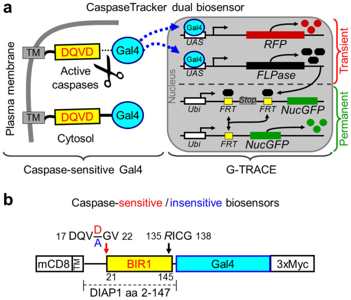
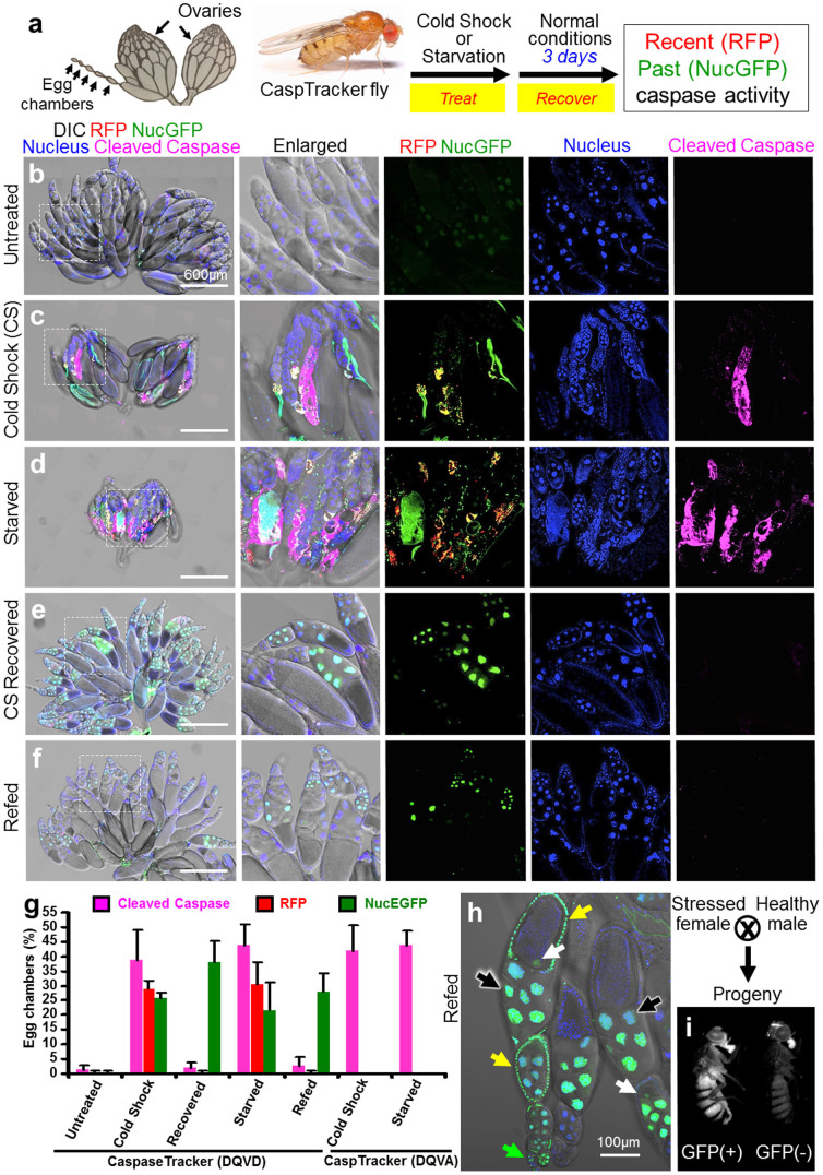
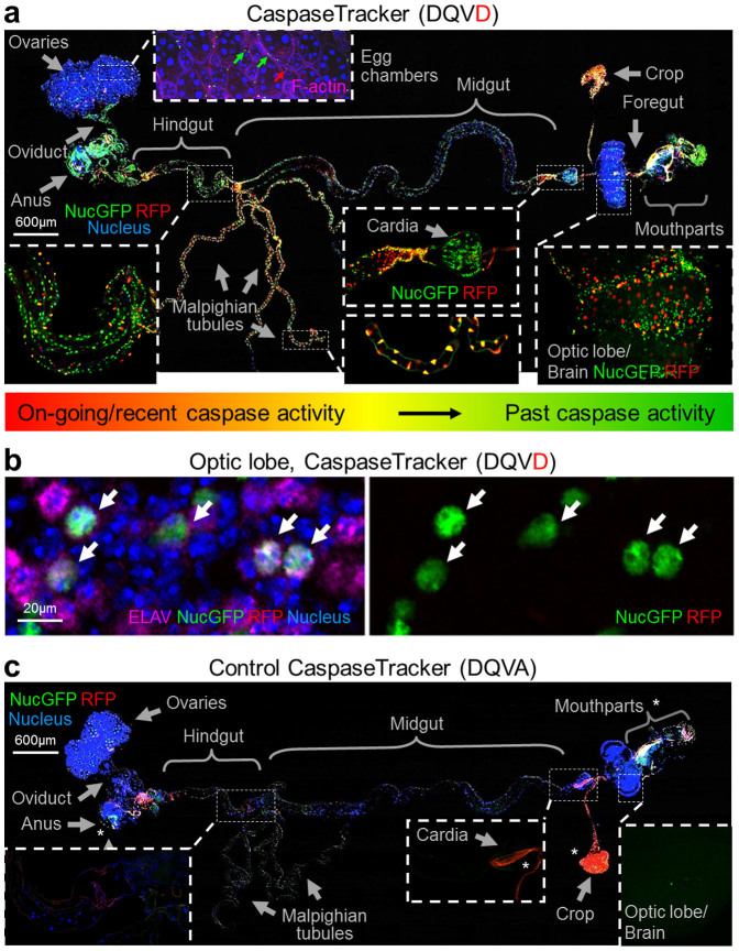
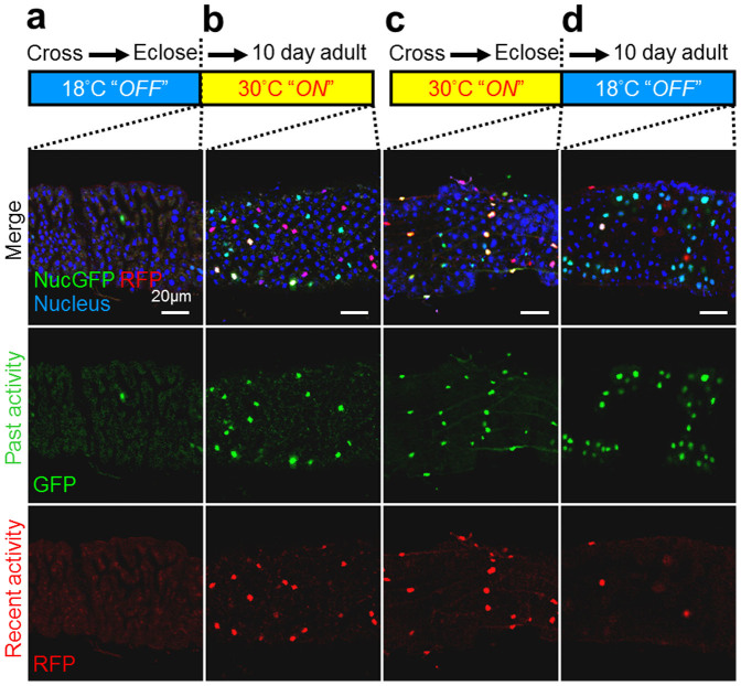
The discovery that mammalian cells can survive late-stage apoptosis challenges the general assumption that active caspases are markers of impending death. However, tools have not been available to track healthy cells that have experienced caspase activity at any time in the past. Therefore, to determine if cells in whole animals can undergo reversal of apoptosis, known as anastasis, we developed a dual color CaspaseTracker system for Drosophila to identify cells with ongoing or past caspase activity. Transient exposure of healthy females to environmental stresses such as cold shock or starvation activated the CaspaseTracker coincident with caspase activity and apoptotic morphologies in multiple cell types of developing egg chambers. Importantly, when stressed flies were returned to normal conditions, morphologically healthy egg chambers and new progeny flies were labeled by the biosensor, suggesting functional recovery from apoptotic caspase activation. In striking contrast to developing egg chambers, which lack basal caspase biosensor activation under normal conditions, many adult tissues of normal healthy flies exhibit robust caspase biosensor activity in a portion of cells, including neurons. The widespread persistence of CaspaseTracker-positivity implies that healthy cells utilize active caspases for non-apoptotic physiological functions during and after normal development.


Figures

Figure 1
Figure 1. CaspaseTracker biosensor system.
(a) Schematic of the CaspaseTracker biosensor system, which is composed of a plasma membrane anchor (mCD8), a caspase-sensitive natural substrate (derived from DIAP1) fused to the Gal4 transcription factor with a C-terminal 3x-myc tag that translocates to the nucleus upon caspase activation to induce the G-Trace system. (b) Schematic of caspase-sensitive (DQVD) and caspase-insensitive control (DQVA) biosensors with the indicated modifications (NN/GV and D135R) to prevent biosensor degradation and to prevent caspase inhibition by the biosensor.
Figure 2
Figure 2. The CaspaseTracker System for detection of apoptosis and anastasis in vivo.
(a) Schematic of Drosophila ovary, and flow chart for cold shock-, and protein starvation-induced cell death in 1-day-old flies, followed by 3-days recovery at normal condition. Drosophila ovary drawing is provided by Polan Santos; Drosophila image is provided by Darren Obbard. Used with permission (b) Egg chambers from the ovary of 6-day female CaspaseTracker flies fed with normal fly food for 6 days (untreated). (c) Caspase biosensor activity in egg chambers of CaspaseTracker Drosophila at 1 day after cold shock (−7°C, 1 hour, followed by 25°C for 24 hours) to induce apoptosis in egg chambers. (d) Caspase biosensor activity in egg chambers of CaspaseTracker Drosophila fed 3 days with 8% sucrose in 1% agar (starved) to induce apoptosis in egg chambers. (e) Like panel c except flies were then switched to normal conditions for 3 days after cold shock (CS recovered). (f) Like panel d except flies were switched to normal yeast-based fly food for 3 days after starvation (refed). Panel at left most is merged confocal image of RFP, NucGFP, nuclei, cleaved-caspase immuno-staining and DIC for overview of egg chambers at the ovaries; middle left panel is enlarged view of the dotted box at the left most panel; middle, middle right, and right most panels display biosensor RFP and NucGFP, nucleus, and cleaved caspase, respectively. (g) Quantification of RFP and NucGFP expression in egg chambers of CaspaseTracker (DQVD) flies before and after apoptosis induction. Caspase insensitive CaspaseTracker (DQVA) files serve as controls. Data presented are from 3 different batches of flies (n = 20), counting 100 egg chambers from each batch per condition. Error bars denote SD. (h) Confocal image of egg chambers recovered 3 days after starvation. Nuclear GFP in nurse cells (black arrows), oocytes (white arrows) and follicle cells (yellow arrows) of egg chambers, and in the germarium (green arrow). (i) GFP and non-GFP expressing progeny from starved and refed CaspaseTracker (DQVD) female flies.
Figure 3
Figure 3
Physiological caspase activity during development (a) Merged confocal image of RFP and NucGFP biosensor, Hoechst for nuclei and DIC of whole mount dissection of newly eclosed day 0 caspase-sensitive CaspaseTracker (DQVD) fly raised at 18°C. Phalloidin for F-actin (pink) is shown only at the sub-panel for egg chambers. (b) NucGFP colocalizes with pan neuronal nuclear immunostaining for ELAV in the optic lobe of CaspaseTracker (DQVD) fly brain. Arrows indicate the co-localized signals of NucGFP and ELAV. (c) Merged confocal image of whole mount dissection of newly eclosed caspase-insensitive CaspaseTracker (DQVA) fly raised at 18°C, imaged with the same condition as panel a. *Autofluorescent regions.
Figure 4
Figure 4. Post developmental caspase activation, and persistence in the adult of cells that experienced developmental caspase activity at Drosophila anterior midgut.
Biosensor fluorescence of anterior midgut of (a) Newly eclosed Gal80ts–temperature sensitive(ts) CaspaseTracker flies raised at 18°C. The thermosensitive Gal80 (Gal80ts) conditionally represses Gal4 at 18°C; biosensor is functional “Off” during development. (b) Newly eclosed 18°C raised (ts) adult CaspaseTracker flies were then shifted to 30°C for 10 days. Gal80ts cannot repress Gal4 at 30°C; biosensor is functional “On” at the 10-day period. (c) Newly eclosed tsCaspaseTracker flies raised at 30°C; biosensor is functional “On” at the development. (d) Newly eclosed 30°C raised (ts) adult CaspaseTracker flies were shifted to 18°C for 10 days. Gal80ts represses Gal4 at 18°C; biosensor is functional “Off” at the 10-day period.

Similar articles

Cited by

References

    1. Alnemri E. S. et al. Human ICE/CED-3 protease nomenclature. Cell 87, 171 (1996). - PubMed
    1. Riedl S. J. & Shi Y. Molecular mechanisms of caspase regulation during apoptosis. Nat Rev Mol Cell Biol 5, 897–907 (2004). - PubMed
    1. Chai J. & Shi Y. Apoptosome and inflammasome: conserved machineries for caspase activation. National Science Review 1, 101–118 (2013).
    1. Jonas E. A. et al. Proapoptotic N-truncated BCL-xL protein activates endogenous mitochondrial channels in living synaptic terminals. Proc Natl Acad Sci U S A 101, 13590–5 (2004). - PMC - PubMed
    1. Li Z. et al. Caspase-3 activation via mitochondria is required for long-term depression and AMPA receptor internalization. Cell 141, 859–71 (2010). - PMC - PubMed

Publication types