. 2015 Mar 11:5:8986.
doi: 10.1038/srep08986.

Prevention of exercised induced cardiomyopathy following Pip-PMO treatment in dystrophic mdx mice

Affiliations

Prevention of exercised induced cardiomyopathy following Pip-PMO treatment in dystrophic mdx mice

Corinne A Betts et al. Sci Rep. .

Abstract

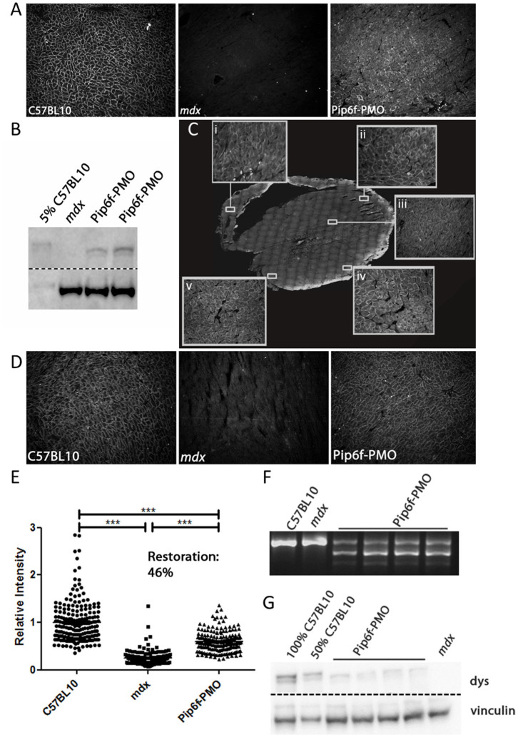
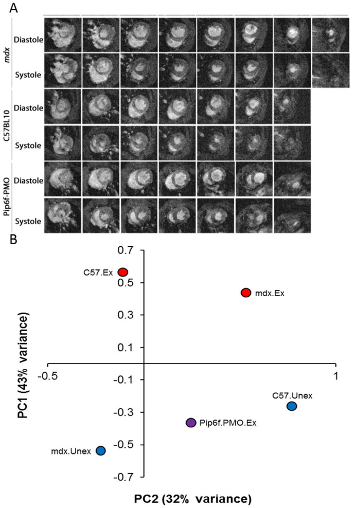
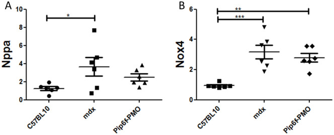
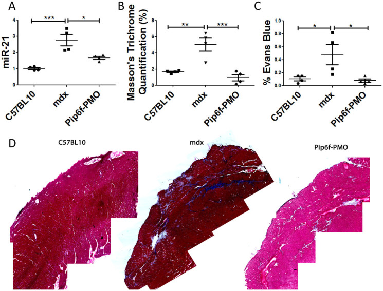
Duchenne muscular dystrophy (DMD) is a fatal neuromuscular disorder caused by mutations in the Dmd gene. In addition to skeletal muscle wasting, DMD patients develop cardiomyopathy, which significantly contributes to mortality. Antisense oligonucleotides (AOs) are a promising DMD therapy, restoring functional dystrophin protein by exon skipping. However, a major limitation with current AOs is the absence of dystrophin correction in heart. Pip peptide-AOs demonstrate high activity in cardiac muscle. To determine their therapeutic value, dystrophic mdx mice were subject to forced exercise to model the DMD cardiac phenotype. Repeated peptide-AO treatments resulted in high levels of cardiac dystrophin protein, which prevented the exercised induced progression of cardiomyopathy, normalising heart size as well as stabilising other cardiac parameters. Treated mice also exhibited significantly reduced cardiac fibrosis and improved sarcolemmal integrity. This work demonstrates that high levels of cardiac dystrophin restored by Pip peptide-AOs prevents further deterioration of cardiomyopathy and pathology following exercise in dystrophic DMD mice.


Conflict of interest statement

A.F.S. and M.J.G. (MRC Technology) and C.A.B., S.M.H. and M.J.A.W. (University of Oxford) are named contributors and benefactors of a patent filed for Pip-PMO technologies described herein. C.A.C., A.M.L.C.S., C.G., G.M., M.A.V., T.C.R. and K.C. declare no competing financial interests

Figures

Figure 1
Figure 1. Dystrophin restoration in heart following Pip6f-PMO treatment in mdx mice.
(A) Representative image of dystrophin immunohistochemical staining in heart following single 10 mg/kg Pip6f-PMO IV administration in mdx mice. (B) Dystrophin western blot of treated mdx hearts relative to 5% C57BL/10 following a single 10 mg/kg Pip6f-PMO administration. Approximately 5% dystrophin restoration observed. All samples run under the same experimental conditions and on the same SDS gel. Dotted line indicates where image was cropped. (C) Mice then underwent 4 daily injections followed by additional administrations every two weeks. Dystrophin immunohistochemical staining of treated heart with inserts indicating higher magnification of designated areas namely the right ventricle (RV) wall (C, i), outer left ventricle (LV) wall at apex (C, ii), inner myocardium (C, iii), and base (C iv and v) of heart. (D) Representative images of immunohistochemical staining of dystrophin protein in exercised C57BL/10, mdx and Pip6f-treated mdx mouse hearts. (E) Quantification of immunohistochemical staining in hearts (from D) following multiple administrations. Dystrophin expression is determined relative to laminin co-stain. The scatter plots show the normalised relative intensity values for each region of interest. Statistical significance was determined using ANOVA followed by Games-Howell post-hoc test to correct for variance heterogeneity (*** = P < 0.001, ** = P < 0.01, * = P < 0.05). (F) Representative images of reverse-transcriptase (RT) PCR illustrating Δ23 splicing in heart Pip6f-PMO treated cohort. All samples run under the same experimental conditions and on the same agarose gel. RT-qPCR was also performed (see Supplementary Fig. 3 B) which resulted in 32.3% Dmd transcripts lacking exon 23, following normalisation to exon 20–21 (SEM 3.1). (G) Representative images of western blots for Pip6f-PMO treated heart. For western blots, 10 μg of protein was loaded and dystrophin (dys) was quantified relative to vinculin loading control. All samples run under the same experimental conditions and on the same SDS gel. Dotted line indicates where image was cropped. For full agarose and SDS gels see Supplementary Fig. 1, 5&6.
Figure 2
Figure 2. Contiguous cine-MRI images and correlation plot of cardiac function parameters in exercised mdx mice following Pip6f-PMO treatment.
(A) Series of contiguous images at 1 mm increments throughout the entire hearts of C57BL/10, mdx and Pip6f-PMO treated mice during diastole and systole (exercised cohorts only). Images displays significantly larger hearts for mdx untreated cohort. (B) Correlation plot of cardiac function parameters measured by cine-MRI in exercised mdx mice following Pip6f-PMO treatment. Principal component analysis plot of all left and right ventricle cardiac function parameters. The two components demonstrate the highest percentage of variance on the Y-axis (component 1, 43%) and X-axis (component 2, 32%). This plot shows how treated mice tend to display values of the component 1 intermediate between C57BL/10 and mdx mice and values of the component 2 corresponding to unexercised mice.
Figure 3
Figure 3. Gene expression analysis for markers of cardiac injury in exercised mdx mice following Pip6f-PMO treatment.
RT-qPCR analysis of Nppa (A) and Nox4 (B) in heart tissue normalised to C57BL/10 cohort. The untreated mdx exercised cohort reveals elevated expression of these injury markers whereas there is partial normalisation for the Pip6f-PMO cohort. Statistical significance was determined using ANOVA followed by Tukey post-hoc test (*** = P < 0.001, ** = P < 0.01, * = P < 0.05).
Figure 4
Figure 4. Reduction in cardiac pathology as determined by the detection of fibrosis and sarcolemmal damage in the hearts of exercised mdx mice following Pip6f-PMO treatment.
(A) RT-qPCR analysis of miRNA-21 in heart tissue normalised to C57BL/10 cohort. (B) Quantification of Masson's trichrome staining in hearts of exercised cohorts. (C) Evans blue dye infiltration into heart following exercise. (D) Masson's trichrome images of worst areas of collagen deposition. miRNA-21 expression and Evans blue dye leakage of Pip6f-PMO hearts is normalised in contrast to untreated control. In addition Masson's trichrome staining is reduced and the representative images indicate less fibrosis then the untreated mdx cohort. Statistical significance was determined using ANOVA followed by Tukey post-hoc test (*** = P < 0.001, ** = P < 0.01, * = P < 0.05).

References

    1. Emery A. E. Population frequencies of inherited neuromuscular diseases--a world survey. Neuromuscul Disord 1, 19–29 (1991). - PubMed
    1. Kirchmann C., Kececioglu D., Korinthenberg R. & Dittrich S. Echocardiographic and electrocardiographic findings of cardiomyopathy in Duchenne and Becker-Kiener muscular dystrophies. Pediatric cardiology 26, 66–72 (2005). - PubMed
    1. Nigro G., Comi L. I., Politano L. & Bain R. J. The incidence and evolution of cardiomyopathy in Duchenne muscular dystrophy. Int J Cardiol 26, 271–277 (1990). - PubMed
    1. Bushby K., Muntoni F. & Bourke J. P. 107th ENMC international workshop: the management of cardiac involvement in muscular dystrophy and myotonic dystrophy. 7th–9th June 2002, Naarden, the Netherlands. Neuromuscul Disord 13, 166–172 (2003). - PubMed
    1. Sultan A. & Fayaz M. Prevalence of cardiomyopathy in Duchenne and Becker's muscular dystrophy. Journal of Ayub Medical College, Abbottabad: JAMC 20, 7–13 (2008). - PubMed

Publication types

MeSH terms