. 2015 Jun;23(6):1055-1065.
doi: 10.1038/mt.2015.41. Epub 2015 Mar 11.

Excision of Expanded GAA Repeats Alleviates the Molecular Phenotype of Friedreich's Ataxia

Affiliations

Excision of Expanded GAA Repeats Alleviates the Molecular Phenotype of Friedreich's Ataxia

Yanjie Li et al. Mol Ther. 2015 Jun.

Abstract

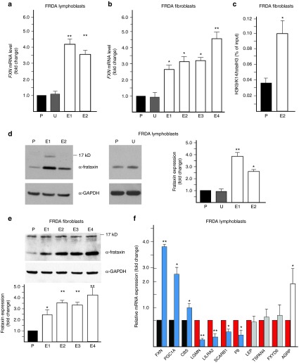
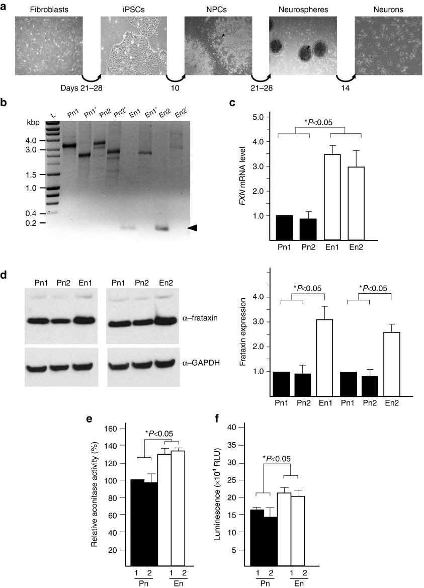
Friedreich's ataxia (FRDA) is an autosomal recessive neurological disease caused by expansions of guanine-adenine-adenine (GAA) repeats in intron 1 of the frataxin (FXN) gene. The expansion results in significantly decreased frataxin expression. We report that human FRDA cells can be corrected by zinc finger nuclease-mediated excision of the expanded GAA repeats. Editing of a single expanded GAA allele created heterozygous, FRDA carrier-like cells and significantly increased frataxin expression. This correction persisted during reprogramming of zinc finger nuclease-edited fibroblasts to induced pluripotent stem cells and subsequent differentiation into neurons. The expression of FRDA biomarkers was normalized in corrected patient cells and disease-associated phenotypes, such as decreases in aconitase activity and intracellular ATP levels, were reversed in zinc finger nuclease corrected neuronal cells. Genetically and phenotypically corrected patient cells represent not only a preferred disease-relevant model system to study pathogenic mechanisms, but also a critical step towards development of cell replacement therapy.


Figures

Figure 1
Figure 1
ZFN targeting of intronic GAA repeats in the FXN gene. (a) Diagram of the FXN gene ZFN editing strategy. Exact location of the UP- and DN-ZFNs is shown relative to the proximal and distal end of the GAA repeat region. Approximate locations of the PCR primers (UP-F/R and DN-F/R) used to amplify ZFN targeted region are indicated. Exact locations and sequences of the primers and ZFNs are shown in Supplementary Figure S1. (b) Simultaneous cleavage by UP- and DN-ZFNs. K562 cells were cotransfected with UP/DN-ZFN mRNAs. In K562 cells harboring the short GAA tract, editing of intron 1 of the FXN gene leads to the release of ~1.2 kbp fragment. Deletion can be detected by PCR using UP-F and DN-R primers. Amplification of the non-GAA edited allele results in a 1.6-kbp fragment (lane C) while amplification of ZFN-edited DNA results in a 0.4-kbp fragment (lane UP-ZFN, DN-ZFN). L—Hyperladder I (BioLine). (c) Schematic illustrating ZFN-mediated editing of the FXN locus.
Figure 2
Figure 2
ZFN-mediated excision of the GAA repeat region in K562 cells does not affect frataxin expression. (a) Analysis of six representative clones derived from K562 cells cotransfected with UP/DN-ZFN mRNAs; N/N designates non-GAA edited clone (~1.6 kbp PCR product), N/E designates heterozygous clones with one edited and one non-GAA edited allele (~1.6 and 0.4 kbp PCR products), and E/E indicates clones with both alleles edited by ZFNs (0.4 kbp PCR product). (b) Results of qRT-PCR analysis of FXN expression. Heterozygous (samples 3, 4, and 6) or homozygous (sample 5) excision of the 1.2-kbp fragment of intron 1 of the FXN gene does not affect expression of FXN mRNA when compared with non-GAA edited cells (samples 1 and 2) or untransfected parental K562 cells (data not shown). Term “non-GAA edited” indicates cells that were nucleofected with UP- and DN-ZFN mRNAs and clonally expanded, but no sequence editing of the GAA region occurred as determined by DNA sequencing analyses of ZFN cleavage regions. Unless otherwise indicated, the non-GAA edited cells were used as controls in all experiments. (c) Editing of the FXN gene does not affect frataxin protein levels in K562 cells as determined by western blot. The asterisk denotes the precursor form of frataxin. Quantitation of the western blot (mature frataxin) is shown below. The results are reported as mean ± SD of three or more experiments. See also Supplementary Figure S6a.
Figure 3
Figure 3
Genetic correction of expanded GAA repeats in FRDA lymphoblasts and fibroblasts. (a) CEL I analysis to determine the efficiency of UP-ZFN cleavage in the heterochromatin region in cells harboring expanded GAAs; FRDA lymphoblasts (GM15850), control lymphoblasts (GM15851); arrowheads indicate expected cleavage products. Quantitative analysis of UP-ZFN activity in FRDA and control lymphoblasts is shown below. WL designates Wide Range DNA marker (Sigma), L—Hyperladder I. (b) Detection of the ZFN-edited alleles in the population of FRDA68 fibroblasts 48 hours post-transfection with UP- and DN-ZFNs (ZFN lane). K562 clone 5 (Figure 2a) was used as a positive control (lane +), whereas a PCR reaction with no template served as a negative control (lane –). (c) Detection of ZFN-edited clones from FRDA lymphoblasts by PCR with UP-F/R primers. Asterisks indicate positive, ZFN-edited clones; C+ positive control (K562 clone 5, Figure 2a), C-negative PCR control. (d) Analysis of the GAA repeat region in two ZFN-edited (E1 and E2) and non-GAA edited clone (P) derived from FRDA lymphoblasts using two PCR primer sets: ZFN-Ext F/R (lanes E1, E2, P) and ZFN-Int (lanes E1′, E2′, and P′) which produce amplicons that differ in the length of the sequences flanking the GAA repeats. The location of all primers relative to the GAA repeats and ZFN cleavage sites is depicted in Supplementary Figure S1. (e) Analysis of the GAA repeat region in identified ZFN-edited and non-GAA edited clones derived from FRDA68 fibroblasts. Clones P, E1, E2, E3, and E4 were analyzed using ZFN-Ext primers; clones P′, E1′, E2′, E3′, and E4′ were amplified with ZFN-Int primers. The short, ~0.2 kbp DNA fragment resulting from ZFN-mediated genetic correction of one of the alleles is indicated by an arrowhead. In some cases, because of the inherent difficulties of amplifying extremely long tracts of GAAs, a long exposure of the gel is required to visualize GAA amplification products (lane E1′). See also Supplementary Figure S6b,c.
Figure 4
Figure 4
Excision of the GAA tract increases frataxin expression and corrects the molecular phenotype of FRDA cells. (a) Determination of FXN mRNA expression using qRT-PCR in the ZFN-edited FRDA lymphoblast clones (E1 and E2) relative to the non-GAA edited control (P) and untransfected control (U). (b) Determination of FXN mRNA expression using qRT-PCR in the ZFN-edited FRDA fibroblast clones (E1–E4) relative to the non-GAA edited control (P) and untransfected control (U). (c) Chromatin immunoprecipitation with an antibody specific to acetylated lysines 9 and 14 on histone H3 (H3K9K14ac) in ZFN-corrected FRDA fibroblast clone E4 and non-GAA edited FRDA fibroblasts (P). (d) Analysis of frataxin expression in ZFN-edited FRDA lymphoblast clones E1 and E2 as determined by western blot. P designates non-GAA edited FRDA lymphoblasts; U designates untransfected FRDA lymphoblasts. Quantitative analysis of frataxin expression is shown in the graph. (e) Analysis of frataxin expression by western blot in the ZFN-corrected FRDA fibroblast clones (E1–E4) relative to the non-GAA edited control (P). Quantitation of the western blot is shown below. (f) The effect of ZFN correction on the FRDA signature of lymphoblast cells. A set of FRDA expression biomarkers was analyzed by qRT-PCR. The expression of the selected mRNAs was normalized to the non-GAA edited FRDA lymphoblasts. The mRNAs known to be underexpressed in FRDA lymphocytes are indicated as black bars while the overexpressed are shown as red bars. Editing of the expanded FXN allele reverses the changes related to frataxin deficiency in six biomarkers (blue bars), does not affect the expression of three mRNAs (gray bars), and has an inverse effect on expression of a single mRNA (white bar). The designations * and ** indicate statistically significant differences with P-values <0.05 and <0.01, respectively. The error bars represent SD of three or more experiments.
Figure 5
Figure 5
Genetic correction of the GAA expansion ameliorates phenotypic defects in iPSC-derived neuronal cells. (a) Schematic illustrating the protocol used to obtain neurons from FRDA fibroblast cells. The arrowheads designate neural rosettes. (b) Analysis of the size of the GAA repeat tract in iPSC-derived neuronal cells using GAA-Int and GAA-Ext PCR primers as described in the legend to Figure 3. The multiple bands visible on the agarose gel result from instability of the GAA repeats during the processes of iPSC reprogramming and differentiation to neuronal cells., Pn1 and Pn2 represent neuronal cells obtained from non-GAA edited iPSCs; En1 and En2 represent neuronal cells obtained from ZFN-edited iPSCs. The ~0.2 kbp DNA fragment resulting from ZFN-mediated genetic correction is indicated by an arrowhead. (c) FXN mRNA expression was determined using qRT-PCR in the corrected neurons (En1 and En2) and noncorrected neurons (Pn1 and Pn2). The data was normalized to the expression of Pn1. (d) Western blot analysis of frataxin levels in Pn1, Pn2, En1, and En2 neurons. (e) Analysis of the aconitase activity in the Pn1, Pn2, En1, and En2 neurons. (f) Intracellular levels of ATP were measured in En1 and En2 neurons and Pn1 and Pn2 neurons. The error bars represent SD of three or more experiments. See also Supplementary Figures S7 and S8.

References

    1. Koeppen, AH (2011). Friedreich's ataxia: pathology, pathogenesis, and molecular genetics. J Neurol Sci 303: 1–12. - PMC - PubMed
    1. Campuzano, V, Montermini, L, Moltò, MD, Pianese, L, Cossée, M, Cavalcanti, F et al. (1996). Friedreich's ataxia: autosomal recessive disease caused by an intronic GAA triplet repeat expansion. Science 271: 1423–1427. - PubMed
    1. Delatycki, MB and Corben, LA (2012). Clinical features of Friedreich ataxia. J Child Neurol 27: 1133–1137. - PMC - PubMed
    1. Herman, D, Jenssen, K, Burnett, R, Soragni, E, Perlman, SL and Gottesfeld, JM (2006). Histone deacetylase inhibitors reverse gene silencing in Friedreich's ataxia. Nat Chem Biol 2: 551–558. - PubMed
    1. Soragni, E, Herman, D, Dent, SY, Gottesfeld, JM, Wells, RD and Napierala, M (2008). Long intronic GAA*TTC repeats induce epigenetic changes and reporter gene silencing in a molecular model of Friedreich ataxia. Nucleic Acids Res 36: 6056–6065. - PMC - PubMed

Publication types

MeSH terms