. 2015 May;56(5):3242-52.
doi: 10.1167/iovs.14-16274.

Lipofuscin redistribution and loss accompanied by cytoskeletal stress in retinal pigment epithelium of eyes with age-related macular degeneration

Affiliations

Lipofuscin redistribution and loss accompanied by cytoskeletal stress in retinal pigment epithelium of eyes with age-related macular degeneration

Thomas Ach et al. Invest Ophthalmol Vis Sci. 2015 May.

Abstract

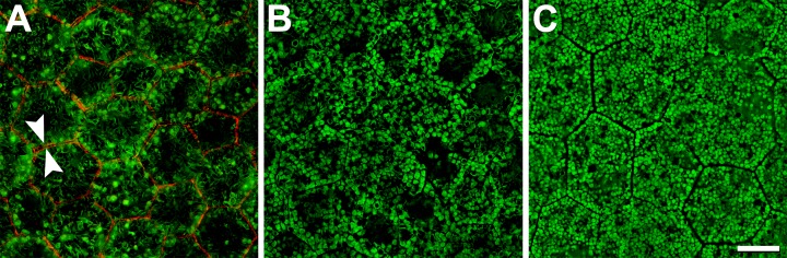
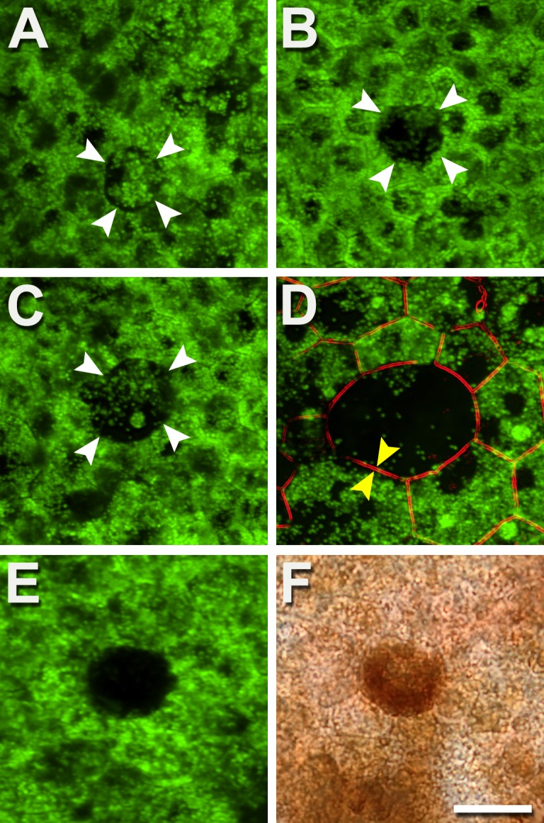
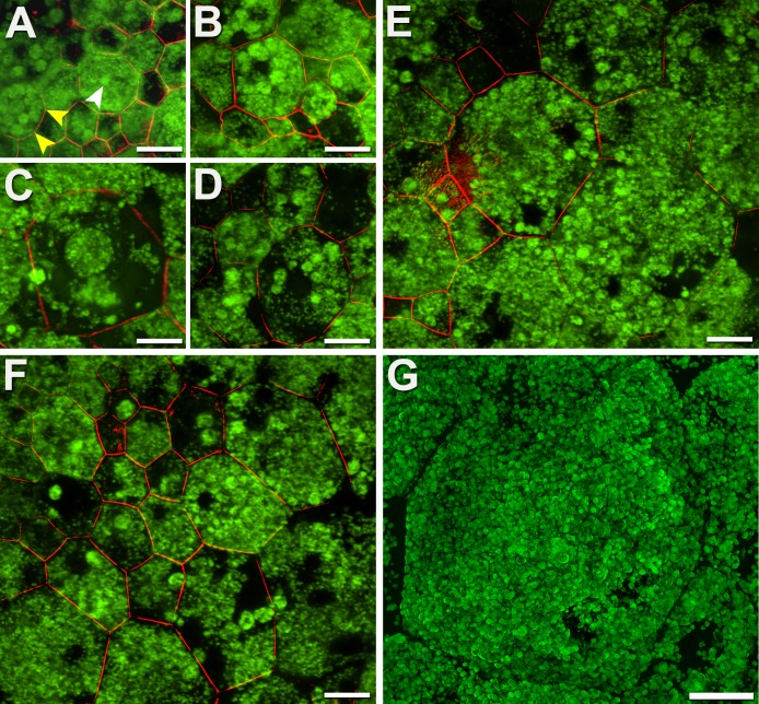
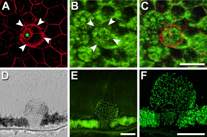
Purpose: Lipofuscin (LF) and melanolipofuscin (MLF) of the retinal pigment epithelium (RPE) are the principal sources of autofluorescence (AF) signals in clinical fundus-AF imaging. Few details about the subcellular distribution of AF organelles in AMD are available. We describe the impact of aging and AMD on RPE morphology revealed by the distribution of AF LF/MLF granules and actin cytoskeleton in human tissues.

Methods: Thirty-five RPE-Bruch's membrane flatmounts from 35 donors were prepared (postmortem: ≤4 hours). Ex vivo fundus examination at the time of accession revealed either absence of chorioretinal pathologies (10 tissues; mean age: 83.0 ± 2.6 years) or stages of AMD (25 tissues; 85.0 ± 5.8 years): early AMD, geographic atrophy, and late exudative AMD. Retinal pigment epithelium cytoskeleton was labeled with AlexaFluor647-Phalloidin. Tissues were imaged on a spinning-disk fluorescence microscope and a high-resolution structured illumination microscope.

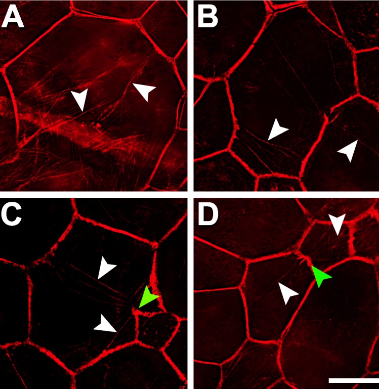
Results: Age-related macular degeneration impacts individual RPE cells by (1) lipofuscin redistribution by (i) degranulation (granule-by-granule loss) and/or (ii) aggregation and apparent shedding into the extracellular space; (2) enlarged RPE cell area and conversion from convex to irregular and sometimes concave polygons; and (3) cytoskeleton derangement including separations and breaks around subretinal deposits, thickening, and stress fibers.

Conclusions: We report an extensive and systematic en face analysis of LF/MLF-AF in AMD eyes. Redistribution and loss of AF granules are among the earliest AMD changes and could reduce fundus AF signal attributable to RPE at these locations. Data can enhance the interpretation of clinical fundus-AF and provide a basis for future quantitative studies.


Figures

Figure 1
Figure 1
Normal RPE granule distribution and F-actin cytoskeleton. High-resolution structured illumination microscopy reveals the intracellular distribution of autofluorescent LF and MLF granules. Melanosomes (minimally AF and not visible) and nonfluorescent cell nuclei lead to dark areas within the cells ([A] fovea; [B] perifovea). With increasing distance from the fovea (out to 10-mm eccentricity), cells contain fewer light-blocking melanosomes and more LF/MLF granules (C). The normal RPE cytoskeleton consists of a circumferential F-actin bundle (red in [A]), which follows the cell's polygonal shape. Adjacent cells have parallel cytoskeletons like railroad tracks (arrowheads in [A]). The F-actin band runs at the apical third of the RPE cell. Retinal pigment epithelial cells can be barrel-shaped and individual cell bodies can thus bulge into adjacent cells basolaterally. From an en face view, granules from one cell might seem to extend into adjacent cells. Donor: 83 years, female. F-actin labeled with AlexaFluor647-Phalloidin. Scale bar: 10 μm.
Figure 2
Figure 2
Autofluorescent granules disappear individually, reducing AF. Degranulation results in a diminished or absent AF signal from affected cells, yet the cells are still present. (A) The central cell (white arrowheads) is circular rather than polygonal and has redistributed granules. (B) The central cell (white arrowheads) is almost devoid of granules. (C) The central cell has a few individual granules and one aggregation (see also Fig. 3). (D) An enlarged cell with a circular profile has an actin cytoskeleton (parallel bands, between yellow arrows) and few granules. (E, F) Epifluorescence and bright-field images of the same area show that decreased AF could also be associated with densely packed melanosomes, in both healthy and AMD eyes. Few AF granules are visible (E). Donors: (AC) 84 years, male, incipient AMD; (D) 86 years, female, incipient AMD; (E, F) 69 years, male, late exudative AMD. Autofluorescent intensity signals were normalized to a fluorescence reference but were not normalized across panels. Scale bar: 20 μm.
Figure 3
Figure 3
Autofluorescent granules aggregate into packets and disappear, reducing AF. (AF) Retinal pigment epithelial cells of AMD eyes sequester LF/MLF granules into dense packets (white arrowhead), in association with a rounding of the cytoskeleton (yellow arrowheads [A]). In late stages, packets of LF/MLF granules are reduced in number diminishing total AF signal further (CF). Aggregation proceeds concurrently with degranulation (D). (G) Massive enlarged cell, possibly healthy, with hundreds of AF granules and not yet undergoing degranulation. A histologic cross-section of granule aggregations is shown in Supplementary Figure S3. Donors: (A, B, DF) 83 years, female, incipient AMD; (C, G) 81 years, male, late nonexudative AMD. (AF) Confocal microscopy, (G) HR-SIM. F-actin labeled with AlexaFluor647-Phalloidin. Scale bars: 20 μm.
Figure 4
Figure 4
Retinal pigment epithelial cells shaped like mushrooms redistribute LF/MLF granules. (AC) En face view: Some RPE cells elevate parts of their cytoplasm into the layer of outer segments. This elevated bleb is delineated by its labeled F-actin (white arrowheads) and exceeds the dimensions of the stem delimited by the cytoskeleton of its neighbors (pentagon; green star in [A]). The bleb is filled with autofluorescent LF/MLF granules (E, F) that when viewed from above has AF signal resembling cells around it (B, C). (DF) Vertical view: A histologic section shows a mushroom-like cell expanding into the layer of outer segments, possibly the initial step of sloughed RPE cells (as described by Zanzottera et al.). Of note, the mushroom cell originates from a continuous RPE layer; microglia, selecting, and engulfing individual RPE cells out of an otherwise intact RPE seems unlikely. Donors: (AC) 69 years, male, atrophic AMD; (DF) 87 years, female, incipient AMD. (A) Fluorescence confocal, F-actin labeled with AlexaFluor647-Phalloidin; (B) fluorescence confocal, 488-nm excitation; (C) image overlay; (D) differential interference contrast mode; (E) fluorescence confocal; (F) fluorescence HR-SIM, both 488-nm excitation. Scale bars: 20 μm.
Figure 5
Figure 5
The cytoskeleton of adjacent RPE cells separates and becomes interrupted. In early stages (A), parallel adjacent cytoskeleton bands are separated partially (≤50%, between white arrowheads). In advanced stages (B, C) greater than 50% cytoskeleton bands are separated. Cytoskeleton interruptions are shown (DI) with free ends, sometimes furled or pointing in different directions. Interrupted cytoskeleton is often associated with AF sub-RPE deposits ([E, H] 488-nm excitation). Donors: (A, B) 94 years, female, incipient AMD; (C) 81 years, male, geographic atrophy; (DI) 86 years, female, late exudative AMD. F-actin labeled with AlexaFluor647-Phalloidin. Scale bars: 20 μm.
Figure 6
Figure 6
Retinal pigment epithelial cells have stress fibers in AMD-affected eyes. Multiple intracellular stress fibers (white arrowheads) arbitrarily cross RPE cells (AD), all of which are enlarged. At sites where stress fibers insert, the cytoskeleton appears frayed and thickened ([C, D] green arrowheads). Although RPE cells are found within atrophic areas, only those outside the atrophic area had recognizable actin cytoskeleton and thus stress fibers. Donors: (A, D) 94 years, female, incipient AMD; (B, C) 81 years, male, geographic atrophy. F-actin labeled with AlexaFluor647-Phalloidin. Scale bar: 20 μm.

References

    1. Delori FC,, Dorey CK,, Staurenghi G,, Arend O,, Goger DG,, Weiter JJ. In vivo fluorescence of the ocular fundus exhibits retinal pigment epithelium lipofuscin characteristics. Invest Ophthalmol Vis Sci. 1995; 36: 718–729. - PubMed
    1. Schmitz-Valckenberg S,, Fleckenstein M,, Scholl HP,, Holz FG. Fundus autofluorescence and progression of age-related macular degeneration. Surv Ophthalmol. 2009; 54: 96–117. - PubMed
    1. von Ruckmann A,, Fitzke FW,, Bird AC. Fundus autofluorescence in age-related macular disease imaged with a laser scanning ophthalmoscope. Invest Ophthalmol Vis Sci. 1997; 38: 478–486. - PubMed
    1. Spaide RF,, Curcio CA. Drusen characterization with multimodal imaging. Retina. 2010; 30: 1441–1454. - PMC - PubMed
    1. Delori FC,, Fleckner MR,, Goger DG,, Weiter JJ,, Dorey CK. Autofluorescence distribution associated with drusen in age-related macular degeneration. Invest Ophthalmol Vis Sci. 2000; 41: 496–504. - PubMed

Publication types