. 2015 Mar 9;32(5):640-51.
doi: 10.1016/j.devcel.2015.01.011.

Cell density sensing alters TGF-β signaling in a cell-type-specific manner, independent from Hippo pathway activation

Affiliations

Cell density sensing alters TGF-β signaling in a cell-type-specific manner, independent from Hippo pathway activation

Flore Nallet-Staub et al. Dev Cell. .

Abstract

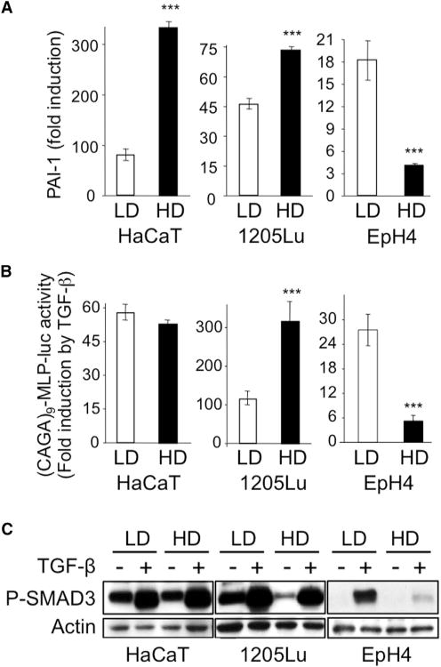
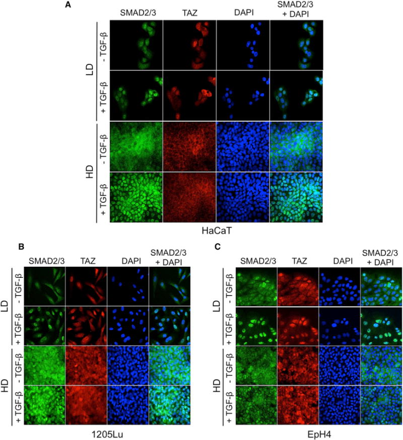
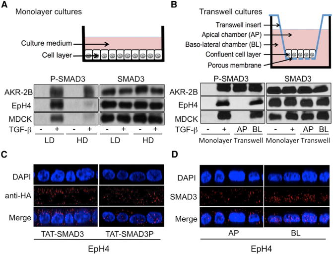
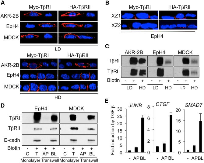
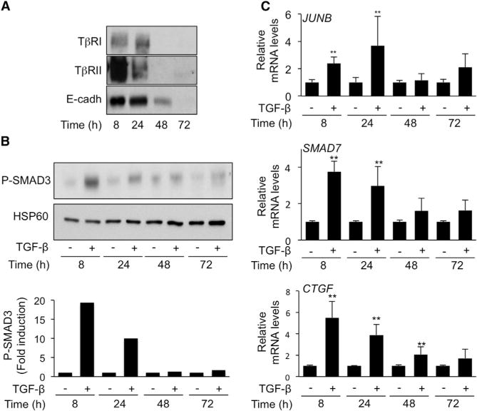
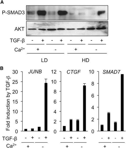
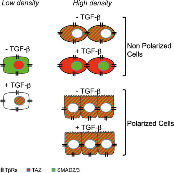
Cell-cell contacts inhibit cell growth and proliferation in part by activating the Hippo pathway that drives the phosphorylation and nuclear exclusion of the transcriptional coactivators YAP and TAZ. Cell density and Hippo signaling have also been reported to block transforming growth factor β (TGF-β) responses, based on the ability of phospho-YAP/TAZ to sequester TGF-β-activated SMAD complexes in the cytoplasm. Herein, we provide evidence that epithelial cell polarization interferes with TGF-β signaling well upstream and independent of cytoplasmic YAP/TAZ. Rather, polarized basolateral presentation of TGF-β receptors I and II deprives apically delivered TGF-β of access to its receptors. Basolateral ligand delivery nonetheless remains entirely effective to induce TGF-β responses. These data demonstrate that cell-type-specific inhibition of TGF-β signaling by cell density is restricted to polarized epithelial cells and reflects the polarized distribution of TGF-β receptors, which thus affects SMAD activation irrespective of Hippo pathway activation.


Figures

Figure 1
Figure 1. Impact of Cell Density on TGF-β Signaling
HaCaT keratinocytes, 1205Lu melanoma cells, and EpH4 mouse mammary epithelial cells were grown in either low (LD) or high (HD) density conditions prior to TGF-β (5 ng/ml) stimulation. (A) Quantitative RT-PCR analysis of PAI-1 expression after a 24-hr TGF-β treatment. Results are expressed as -fold induction by TGF-β in each culture condition and are the mean ± SD from three independent experiments, each measured in triplicate. (B) Effect of TGF-β on SMAD3/4-specific transcription. Results are expressed as -fold activation of transiently transfected (CAGA)9-MLP-luc activity 18 hr after TGF-β addition to the cultures. Results are the mean ± SD of two independent experiments, each performed with triplicate samples. (C) Western analysis of P-SMAD3 levels without or with 30 min TGF-β stimulation. Actin levels were measured as a control for the specificity of P-SMAD3 changes under each experimental condition. Results from one representative of several independent experiments are shown.
Figure 2
Figure 2. Independent Regulation of SMAD2/3 Nuclear Translocation and Density-Dependent TAZ Nucleo-Cytoplasmic Localization
(A–C) Simultaneous detection of SMAD2/3 and TAZ in HaCaT keratinocytes (A), 1205Lu melanoma cells (B), and EpH4 mouse mammary epithelial cells (C). Cells were grown on glass coverslips in either low (LD) or high (HD) density conditions prior to TGF-β (30 min, 5 ng/ml) stimulation, then subjected to simultaneous immunofluorescent detection of SMAD2/3 (green) and TAZ (red). Experiments were repeated four to six times, depending on cell line.
Figure 3
Figure 3. Plasma Membrane Receptor Localization Determines Cell- and Density-Specific TGF-p Responsiveness
(A) Western analysis of P-SMAD3 (left) and SMAD3 (right) levels in AKR-2B fibroblasts, EpH4, and MDCK epithelial cells grown at low (LD) or high (HD) density monolayer cultures (schematic) following a 30-min TGF-β stimulation. (B) AKR-2B, MDCK, and EpH4 cells were grown in monolayer (at LD) or Transwell cultures (schematic). P-SMAD3 (left) and SMAD3 levels (right) were determined by western blotting in monolayer cultures following 60 min stimulation in the absence (−) or presence (+) of TGF-β or subsequent to Transwell apical (AP) or basolateral (BL) TGF-β addition. (C) Confluent monolayers of EpH4 cells were transduced with either TAT-Smad3 (left) or TAT-Smad3P (right). Forty-five minutes later, cells were fixed and subcellular localization of TAT-Smad3 and TAT-Smad3P was assessed by confocal microscopy. Representative results are shown. Nuclei (blue) were stained with DAPI. (D) Immunofluorescent detection of SMAD3 in polarized EpH4 cells by confocal microscopy following a 30min incubation with TGF-β added either to the apical (AP, left) or basolateral (BL, right) Transwell chamber.
Figure 4
Figure 4. Loss of Apical TGF-β Responsiveness in Polarized Epithelial Cells Reflects Exclusive Basolateral TGF-β Receptor Expression
(A) IF detection of transiently transfected Myc-TβRI and HA-TβRII in low density (LD, upper) and high density (HD, lower) monolayer cultures of AKR-2B, MDCK, and EpH4 cells. Images are represented as XZ cross-sections of transfected cells. (B) IF detection of transiently transfected Myc-TβRI and HA-TβRII in polarized Transwell cultures of EpH4 cells. Two representative XZ cross-sections of transfected cells are shown. Nuclei (blue) were stained with DAPI. (C) Surface biotinylation assays of endogenous TβRI and TβRII in AKR-2B, EpH4, and MDCK cells grown in low- or high-density monolayer cultures. (D) Apical (AP) and basolateral (BL) surface biotinylation assays of endogenous TβRI and TβRII in MDCK and EpH4 cells in monolayer (low-density) or Transwell cultures (high density, polarized). Cell proteins were biotinylated either from the medium (monolayer on plastic, low density) or from the apical or basolateral medium in the case of high density polarized transwell cultures. After surface biotinylation, proteins were immunoprecipitated with streptavidin-agarose and then detected by blotting with the appropriate TGF-β receptor or E cadherin antibody. Lanes C and T reflect immunoprecipitated protein in the absence of biotinylation (C) or in monolayer for total labeling (T), respectively. E-cadherin was detected to serve as a marker of basolateral cell surfaces. (E) Quantitative reverse transcriptase–PCR analysis of JUNB, CTGF, and SMAD7 expression in polarized EpH4 cells incubated without (−) or with TGF-β (2 hr) added either in the apical (AP) or basolateral (BL) compartment of Transwells. Results are expressed as -fold induction by TGF-β and are the mean ± SD from two independent experiments, each measured in triplicate.
Figure 5
Figure 5. TβR Basolateral Relocalization during EpH4 Cell Polarization: Implication for TGF-β Signaling
EpH4 mouse mammary epithelial cells were grown in high-density conditions (see Experimental Procedures). (A) Surface biotinylation assays of endogenous TβRI, TβRII, and E-cadherin performed at various time points following cell seeding. One of two experiments with sub-identical results is shown. (B) Western analysis of P-SMAD3 levels following a 2-hr TGF-β stimulation at various time points following cell seeding. Upper panel: representative blot from one of five experiments that gave similar results. Lower: Densitometric analysis of P-SMAD3 levels. Results are expressed as -fold induction by TGF-β at each time-point. (C) Quantitative reverse transcriptase–PCR analysis of JUNB, CTGF, and SMAD7 expression in EpH4 cells incubated without (−) or 2 hr with TGF-β at various time points following cell seeding. Results, expressed as -fold induction by TGF-β at each time point are the mean ± SD from five independent experiments, each measured in triplicate.
Figure 6
Figure 6. Effect of Ca2+ Depletion on TGF-β Response in High-Density Monolayer Cultures of EpH4 Cells
(A) Western analysis of P-SMAD3 levels in EpH4 cells treated without (−) or with (+) TGF-β for 30 minat low (LD) or high (HD) density in monolayer cultures in the presence (+) or absence (−) of Ca2+. (B) Quantitative reverse transcriptase-PCR analysis of JUNB, CTGF, and SMAD7 expression in confluent monolayers of EpH4 cells incubated without (−) or with TGF-β (2 hr) in medium with (+) or depleted of (−) Ca2+. Results are expressed as -fold induction by TGF-β and are the mean ± SD from two independent experiments, each measured in triplicate.
Figure 7
Figure 7. Schematic Summary of TAZ and SMAD3 Nucleo-Cytoplasmic Shuttling in Response to Cell Density or TGF-β Stimulation in Non-Polarized or Polarized Cells at Low or High Density
At low density (LD), TAZ (red) is nuclear and SMAD3 (green) is cytoplasmic. TGF-β receptors are distributed throughout the cell surface. In response to TGF-β, SMAD3 accumulates in the nucleus where TAZ is located. At high density (HD), TAZ is excluded from the nucleus, as a result of cell density sensing. In non-polarized cells, TGF-β induces SMAD3 nuclear accumulation, while TAZ remains cytoplasmic. In polarized cells (schematically represented with apical villi), apical delivery of TGF-β is without effect on SMAD localization because TβRs are exclusively basolateral and TGF-β access to its receptors is blocked by intercellular junctions.

References

    1. Alarcón C, Zaromytidou AI, Xi Q, Gao S, Yu J, Fujisawa S, Barlas A, Miller AN, Manova-Todorova K, Macias MJ, et al. Nuclear CDKs drive Smad transcriptional activation and turnover in BMP and TGF-beta pathways. Cell. 2009;139:757–769. - PMC - PubMed
    1. Alexaki VI, Javelaud D, Mauviel A. JNK supports survival in melanoma cells by controlling cell cycle arrest and apoptosis. Pigment Cell Melanoma Res. 2008;21:429–438. - PubMed
    1. Alexaki VI, Javelaud D, Van Kempen LC, Mohammad KS, Dennler S, Luciani F, Hoek KS, Juàrez P, Goydos JS, Fournier PJ, et al. GLI2-mediated melanoma invasion and metastasis. J Natl Cancer Inst. 2010;102:1148–1159. - PMC - PubMed
    1. Beyer TA, Weiss A, Khomchuk Y, Huang K, Ogunjimi AA, Varelas X, Wrana JL. Switch enhancers interpret TGF-β and Hippo signaling to control cell fate in human embryonic stem cells. Cell Rep. 2013;5:1611–1624. - PubMed
    1. Chen X, Xu H, Yuan P, Fang F, Huss M, Vega VB, Wong E, Orlov YL, Zhang W, Jiang J, et al. Integration of external signaling pathways with the core transcriptional network in embryonic stem cells. Cell. 2008;133:1106–1117. - PubMed

Publication types

MeSH terms

LinkOut - more resources