. 2015 Oct;40(10):2091-101.
doi: 10.1007/s11064-015-1548-7. Epub 2015 Mar 11.

Paraoxon and Pyridostigmine Interfere with Neural Stem Cell Differentiation

Affiliations

Paraoxon and Pyridostigmine Interfere with Neural Stem Cell Differentiation

Verónica O Berríos et al. Neurochem Res. 2015 Oct.

Abstract

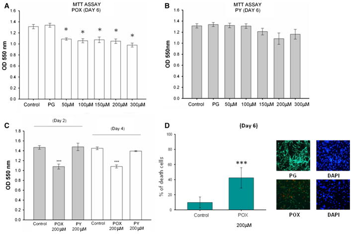
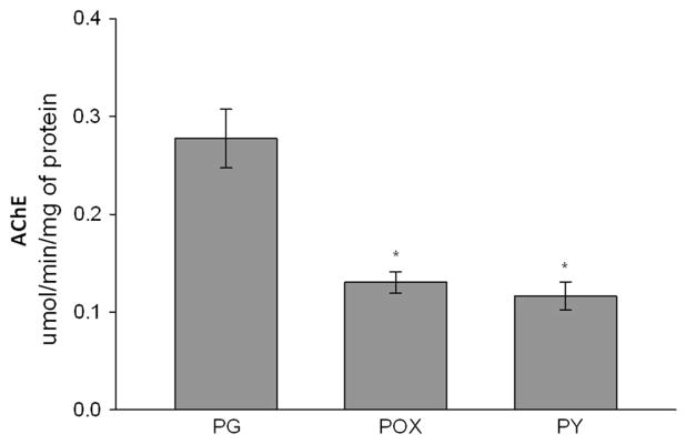
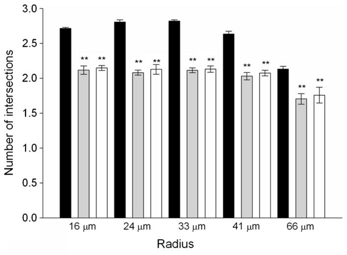
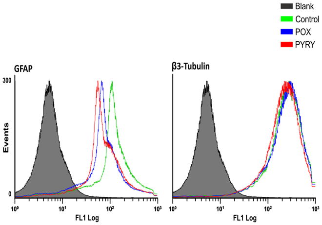
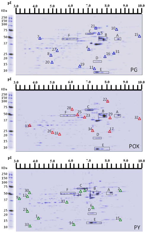
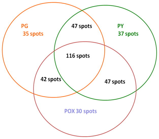
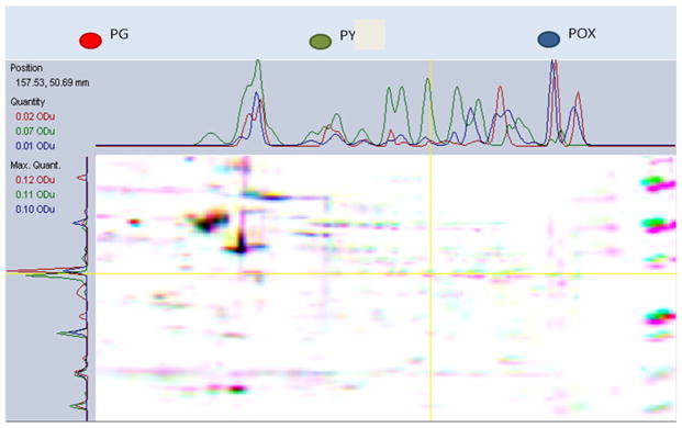
Acetylcholinesterase (AChE) inhibition has been described as the main mechanism of organophosphate (OP)-evoked toxicity. OPs represent a human health threat, because chronic exposure to low doses can damage the developing brain, and acute exposure can produce long-lasting damage to adult brains, despite post-exposure medical countermeasures. Although the main mechanism of OP toxicity is AChE inhibition, several lines of evidence suggest that OPs also act by other mechanisms. We hypothesized that rat neural progenitor cells extracted on embryonic day 14.5 would be affected by constant inhibition of AChE from chronic exposure to OP or pyridostigmine (a reversible AChE blocker) during differentiation. In this work, the OP paraoxon decreased cell viability in concentrations >50 μM, as measured with the MTT assay; however, this effect was not dose-dependent. Reduced viability could not be attributed to blockade of AChE activity, since treatment with 200 µM pyridostigmine did not affect cell viability, even after 6 days. Although changes in protein expression patterns were noted in both treatments, the distribution of differentiated phenotypes, such as the percentages of neurons and glial cells, was not altered, as determined by flow cytometry. Since paraoxon and pyridostigmine each decreased neurite outgrowth (but did not prevent differentiation), we infer that developmental patterns may have been affected.

Keywords: Differentiation; Neural progenitor cells; Organophosphates; Paraoxon; Proteomics.


Conflict of interest statement

Conflict of interest The authors declare that they have no conflict of interest.

Figures

Fig. 1
Fig. 1
Interference of paraoxon (POX) and pyridostigmine (PY) with cell viability. MTT assay of differentiating neural progenitor cells treated with POX (a) or PY (b) for 6 days (treated cells vs. PG treatment *p <0.05). c MTT assay of differentiating neural progenitor cells treated with POX or PY and analyzed on days 2 and 4 (***p <0.001). d Representative immunocytochemistry using antibodies against β3-tubulin and a secondary antibody conjugated with Alexa Fluor® 488 (green), 5 μM propidium iodide (red), and 1 μg/ml of 4′,6-diamidino-2-phenylindole (DAPI, blue). Left panel quantification of the number of propidium iodide-positive cells (p <0.001). Percentage of cell death determined by the intensity ratio propidium iodide/DAPI. Values are the mean number of cells in three independent experiments. Statistical analysis was performed using one-way ANOVA followed by the Tukey test (a, b, c) or by Student’s t-test (d)
Fig. 2
Fig. 2
AChE activity in differentiating neural progenitor cells in the presence of propylene glycol (PG), paraoxon (POX), or pyridostig-mine (PY). The cells were harvested on day 6 after treatment. AChE activity was measured using the Ellman assay, and the results were as follows: PG, 0.28 ± 0.03 (mean ± S.E.M.), n = 6; POX, 0.13 ± 0.01, n = 6; PY, 0.12 ± 0.015, n = 9. *p < 0.05. Statistical analysis was performed using one-way ANOVA followed by the Student–Newman–Keuls test
Fig. 3
Fig. 3
Paraoxon decreases the number of neurite per neuron after 6 days of treatment with either POX or PY. Morphological (Sholl) analysis was performed on differentiating neural progenitor cells stained with an antibody against β3-tubulin. **p < 0.01. Statistical analysis was performed using one-way ANOVA followed by the Tukey test, n = 60, for each treatment. Black bar control, Gray POX, White PY
Fig. 4
Fig. 4
Maintenance of cellular fate decision of differentiating neural progenitor cells in the presence of paraoxon (POX), pyridostigmine (PY), or the vehicle control propylene glycol (PG). Flow cytometry analysis using antibodies against GFAP and β3-tubulin after 6 days of differentiation with different treatments. The graphs shown are one-parameter histograms with cell counts on the y-axis and the measurement parameter on the x-axis. Blank cells, without primary antibodies and with control-only secondary antibody. There were no significant differences between the control-, PY-, and POX-treated groups
Fig. 5
Fig. 5
Overview of two-dimensional gel electrophoresis (2D-GE) showing protein profiles in differentiating neural progenitor cells in the presence of propylene glycol (PG) vehicle, paraoxon (POX), or pyridostigmine (PY). The blue triangles represent differentially expressed proteins in comparison with POX and PY; the red triangles represent the differentially expressed proteins following POX treatment; and the green triangles represent the differentially expressed proteins following PY treatment in comparison with PG. The spots on the 2D gels that are common to all the three conditions are indicated with capital letters. All experiments were performed in triplicate
Fig. 6
Fig. 6
Venn diagram of common and uniquely expressed proteins in differentiating neural progenitor cells cultured in the presence of propylene glycol (PG), paraoxon (POX), or pyridostigmine (PY)
Fig. 7
Fig. 7
Multichannel viewer highlighting the overlay of three 2D gels of neural progenitor cell proteins in the presence of propylene glycol (PG), paraoxon (POX), or pyridostigmine (PY). The images depict a computer-generated, color-coded comparison between the three samples and exhibit intensity variations in the x- and y-directions

Similar articles

Cited by

References

    1. Casida JE, Quistad GB. Organophosphate toxicology: safety aspects of nonacetylcholinesterase secondary targets. Chem Res Toxicol. 2004;17(8):983–998. - PubMed
    1. Main AR. Kinetics of cholinesterase inhibition by organophosphate and carbamate insecticides. Can Med Assoc J. 1969;100(4):161–167. - PMC - PubMed
    1. Andersen JB, et al. Total drug treatment and comorbidity in myasthenia gravis: a population-based cohort study. Eur J Neurol. 2014;21(7):948–955. - PMC - PubMed
    1. Hail SL, Obafemi A, Kleinschmidt KC. Successful management of olanzapine-induced anticholinergic agitation and delirium with a continuous intravenous infusion of physostigmine in a pediatric patient. Clin Toxicol (Phila) 2013;51(3):162–166. - PubMed
    1. Butler AM, Murray M. Biotransformation of parathion in human liver: participation of CYP3A4 and its inactivation during mi-crosomal parathion oxidation. J Pharmacol Exp Ther. 1997;280(2):966–973. - PubMed

Publication types

LinkOut - more resources