. 2015 May;47(5):177-86.
doi: 10.1152/physiolgenomics.00138.2014. Epub 2015 Mar 10.

Similarities and differences of X and Y chromosome homologous genes, SRY and SOX3, in regulating the renin-angiotensin system promoters

Affiliations

Similarities and differences of X and Y chromosome homologous genes, SRY and SOX3, in regulating the renin-angiotensin system promoters

Fabiano C Araujo et al. Physiol Genomics. 2015 May.

Abstract

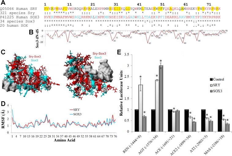
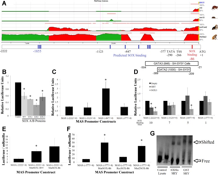
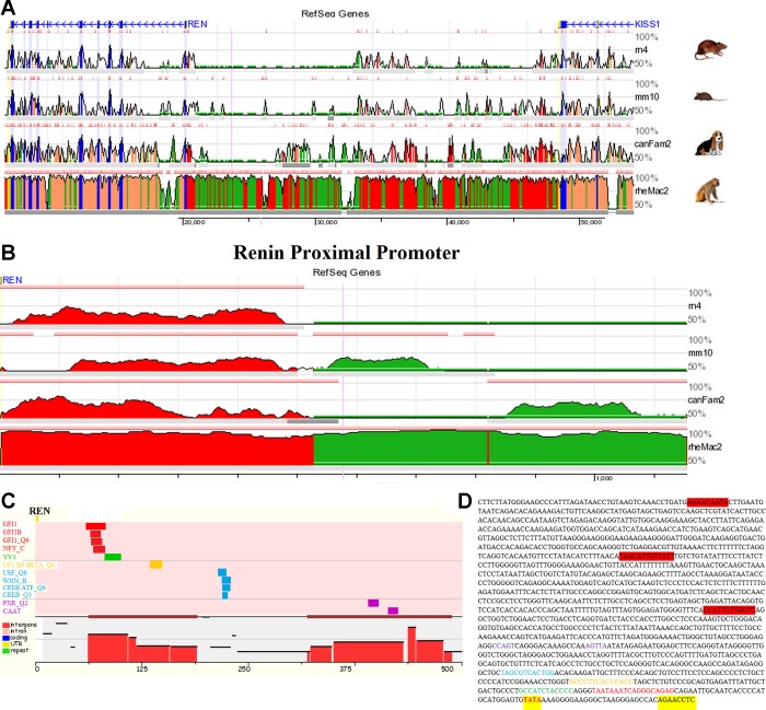
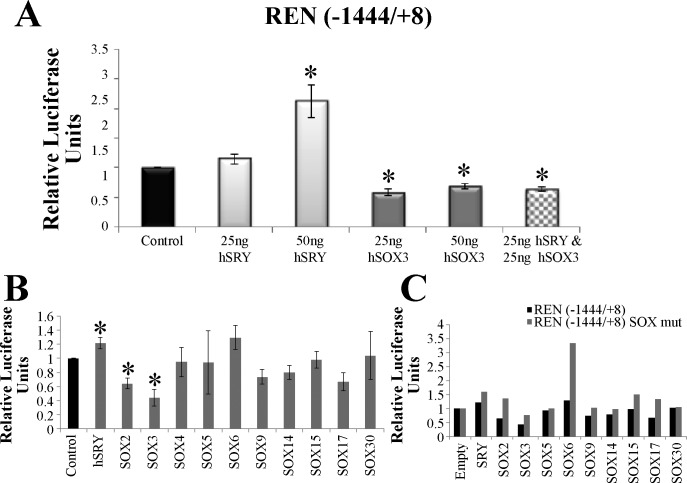
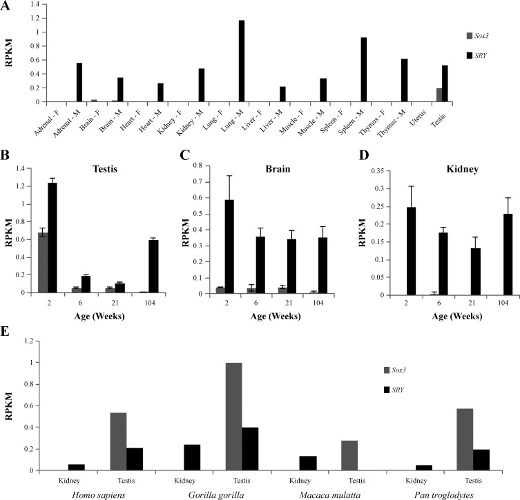
The renin-angiotensin system (RAS) is subject to sex-specific modulation by hormones and gene products. However, sex differences in the balance between the vasoconstrictor/proliferative ACE/ANG II/AT1 axis, and the vasodilator/antiproliferative ACE2/ANG-(1-7)/MAS axis are poorly known. Data in the rat have suggested the male-specific Y-chromosome gene Sry to contribute to balance between these two axes, but why the testis-determining gene has these functions remains unknown. A combination of in silico genetic/protein comparisons, functional luciferase assays for promoters of the human RAS, and RNA-Seq profiling in rat were used to address if regulation of Sry on the RAS is conserved in the homologous X-chromosome gene, Sox3. Both SRY and SOX3 upregulated the promoter of Angiotensinogen (AGT) and downregulated the promoters of ACE2, AT2, and MAS, likely through overlapping mechanisms. The regulation by both SRY and SOX3 on the MAS promoter indicates a cis regulation through multiple SOX binding sites. The Renin (REN) promoter is upregulated by SRY and downregulated by SOX3, likely through trans and cis mechanisms, respectively. Sry transcripts are found in all analyzed male rat tissues including the kidney, while Sox3 transcripts are found only in the brain and testis, suggesting that the primary tissue for renin production (kidney) can only be regulated by SRY and not SOX3. These results suggest that SRY regulation of the RAS is partially shared with its X-chromosome homolog SOX3, but SRY gained a sex-specific control in the kidney for the rate-limiting step of the RAS, potentially resulting in male-specific blood pressure regulation.

Keywords: Sox3; Sry; hypertension; renin angiotensin system; sex differences.


Figures

Fig. 1.
Fig. 1.
Functional conservation in SRY and SOX3. A: sequence alignment of the high-mobility group (HMG) box of human SOX3 and SRY with conservation shown below each in multiple species. Those sites marked with an asterisk (*) are 100% conserved in all species and those with a colon (:) are functionally conserved. Amino acids in red are conserved in all SRY and SOX3, while those in cyan are conserved in SOX3 only. Amino acids highlighted in yellow are those in which mutations in SRY are associated with sex reversal (50). B: normalized values for each amino acid's (x-axis) nonsynonymous rate (dN) minus the synonymous rate (dS) for the 34 sequences of Sox3. Values more negative suggest codon selection. C: known structure of SRY bound to DNA (pdb file 1j46) with the amino acids conserved among all SRY and SOX3 sequences shown in red and those conserved in SOX3 but not SRY shown in cyan. DNA is shown in gray. D: root-mean squared fluctuation (RMSF) for each amino acid over a 3.25 nanosecond molecular dynamic simulation for the known structure of SRY (red) or the modeled SOX3 (cyan) indicate similarities of the 2 proteins. E: the pEF1 expression vectors for either human SRY (light gray) or SOX3 (darker gray) were transfected into CHO cells along with a pGL3 vector containing the human promoter (shown on the bottom) of various RAS genes driving the production of luciferase. Each sample was normalized to an empty vector control of pEF1 (black). Error bars are shown as the SE. Each sample is compared with the control for each promoter. *Significant differences (P ≤ 0.05).
Fig. 2.
Fig. 2.
Functional SOX binding sites in the MAS promoter. A: evolutionary conserved region (ECR) browser analysis of the MAS pGL3 promoter construct for the rat (rn4), mouse (mm10), dog (canFam2), and monkey (rh2Mac2). Regions in red and green share conservation with human based on 20 bp stretches sharing >50% homology. Below the ECR is the location of various landmarks of the promoter vector such as sites used to create cleavage series (−2222, −1123, and −377), predicted SOX binding sites (blue), in vitro confirmed SOX binding site (red), the location of the TATA box (−290), proposed transcriptional start site (TSS, −266), translational start site (ATG, +1), and ENCODE transcription factor binding with relative score and cell line of observation. B: the human MAS promoter treated with various SOX A/B proteins, all of which significantly repress the promoter. C: luciferase production of 4 MAS promoter constructs with differing lengths, all made relative to the full-length MAS (−2222/+4) construct regulation level. D: promoter analysis of 4 MAS promoter constructs transfected with control (black), SRY (light gray) or SOX3 (dark gray) vectors. Total number of SOX binding sites in each construct is shown below the graph. E: mutations to the MAS(−2222/4) promoter construct at 2 of the 10 SOX binding sites (−1855 or −86) result in elevation of promoter activity. Data are shown as the raw luciferase-Renilla ratio. F: as the MAS(−2222/+4)MutSOX-86 showed a trend in altering regulation and was previously shown to be an in vitro binding site for SRY, the site was investigated with the smaller MAS minimal promoter, MAS(−377/+4). The SOX binding site was mutated to 2 separate sequences, MAS(−377/+4)MutSOX-86 and MAS(−377/+4)Mut2SOX-86, resulting in elevation of promoter activity. One of the mutations was repaired to recover the SOX binding site, MAS(−377/+4)RecSOX-86, confirming only this site was altered in the promoter mutagenesis. G: the sequence for the SOX binding site at −86 was synthesized with a biotin tag and EMSA performed using both His- and GST-tagged Sry proteins, confirming that SOX proteins can bind to this element. Error bars for all graphs are shown as the SE. Each sample is compared with the control for each promoter. *Significant differences (P ≤ 0.05) relative to the control.
Fig. 3.
Fig. 3.
Similar mechanisms of regulation by SRY and SOX3 on promoters of AGT and ACE2. A: activation of the AGT promoter with 2 concentrations of transfected SRY (light gray) or SOX3 (dark gray). In addition, a double transfection of SRY and SOX3 (checkered) shows nonadditive activation suggesting shared mechanisms of promoter control. B: repression of the ACE2 promoter with 2 concentrations of transfected SRY (light gray) or SOX3 (dark gray). In addition a double transfection of SRY and SOX3 (checkered) shows nonadditive repression, suggesting shared mechanisms of promoter control. Error bars are shown as the SE. Each sample is compared with the control for each promoter. *Significant differences (P ≤ 0.05).
Fig. 4.
Fig. 4.
Promoter analysis of the gene renin (Ren). A: ECR browser analysis of the intergenic space (roughly 30 kB) between Renin (REN) and the syntenic KiSS-1 metastasis-suppressor gene (KISS1) for the rat (rn4), mouse (mm10), dog (canFam2), and monkey (rh2Mac2). Regions in blue are protein coding exons, yellow are untranslated regions (UTRs), and those in red and green share conservation with human based on 20 bp stretches sharing ≥ 90% homology. B: ECR browser analysis for the proximal promoter of REN with regions in red and green showing conservation of 50 bp stretches with 50% homology. C: the large ECR in the proximal promoter of REN was analyzed for shared transcription factor (TF) binding sites between human and rat using the RVISTA software selecting for all possible TFs. D: the sequence of REN(−1444/+8) pGL3 promoter construct identifying the TATA box (red, highlighted yellow) located within the proper distance of the 5′-UTR (highlighted yellow). Additionally the conserved TF sites are colored relative to that in C. The 3 potential SOX binding sites (highlighted in red) fall outside of the ECR.
Fig. 5.
Fig. 5.
Regulation of the REN promoter by various SOX proteins. A: activation of the REN promoter with 2 concentrations of transfected SRY (light gray) or repression by 2 concentrations of SOX3 (dark gray). In addition a double transfection of SRY and SOX3 (checkered) showed downregulation of the promoter similar to that of SOX3 transfected cells. B: transfection of CHO cells with either the control pEF1 vector (black) or 1 of the SOX pEF1 constructs (gray) along with the pGL3 REN(−1444/+8) luciferase vector. Error bars are shown as the SE. Each sample is compared with the control for each promoter. *Significant differences (P ≤ 0.05). C: the REN(−1444/+8) promoter construct was mutated to remove each of the 3 SOX binding sites; this construct was named REN(−1444/+8) SOX mut. The mutant pGL3 construct (gray) was transfected with each of the SOX pEF1 expression vectors resulting in an increase in promoter activity relative to the wt pGL3 promoter construct (black) for all but the SOX30 vector (n = 1).
Fig. 6.
Fig. 6.
Expression profile for Sry and Sox3 in the rat using data from the Rat BodyMap project. Expression profile for Sox3 (gray) and Sry (black) in female (F) and male (M) RNA-Seq data for 10 tissues in the Fisher 344 rat (A). Each tissue is a pool of 16 individual RNA-Seq experiments (4 animals at 4 ages). Data are shown as reads per kilobase per million (RPKM). While Sry is ubiquitously expressed in male only, Sox3 transcripts are found at all 4 ages (2, 6 21, and 104 wk old rats) in the male testis (B) and brain (C). The kidney showed expression of Sry at all 4 ages with no detectable Sox3 reads (D). Error bars for the individual tissues (B–D) represent the SE of 4 independent RNA-Seq datasets at each age. E: analysis of primate (Homo sapiens, Gorilla gorilla, Macaca mulatta, Pan troglodytes) kidney and testis SRA datasets for expression of SOX3 (gray) or SRY (black). Expression of both SRY and SOX3 are seen in testis, while only SRY expression is found in kidney.

References

    1. Alatzoglou KS, Kelberman D, Dattani MT. The role of SOX proteins in normal pituitary development. J Endocrinol 200: 245–258, 2009. - PubMed
    1. Altschul SF, Gish W, Miller W, Myers EW, Lipman DJ. Basic local alignment search tool. J Mol Biol 215: 403–410, 1990. - PubMed
    1. Bellott DW, Hughes JF, Skaletsky H, Brown LG, Pyntikova T, Cho TJ, Koutseva N, Zaghlul S, Graves T, Rock S, Kremitzki C, Fulton RS, Dugan S, Ding Y, Morton D, Khan Z, Lewis L, Buhay C, Wang Q, Watt J, Holder M, Lee S, Nazareth L, Rozen S, Muzny DM, Warren WC, Gibbs RA, Wilson RK, Page DC. Mammalian Y chromosomes retain widely expressed dosage-sensitive regulators. Nature 508: 494–499, 2014. - PMC - PubMed
    1. Berta P, Hawkins JB, Sinclair AH, Taylor A, Griffiths BL, Goodfellow PN, Fellous M. Genetic evidence equating SRY and the testis-determining factor. Nature 348: 448–450, 1990. - PubMed
    1. Bodiga VL, Bodiga S. Renin angiotensin system in cognitive function and dementia. Asian J Neurosci 2013: e102602, 2013.

Publication types

MeSH terms

LinkOut - more resources