. 2015 Mar 10;6(2):e00133.
doi: 10.1128/mBio.00133-15.

A group A Streptococcus ADP-ribosyltransferase toxin stimulates a protective interleukin 1β-dependent macrophage immune response

Affiliations

A group A Streptococcus ADP-ribosyltransferase toxin stimulates a protective interleukin 1β-dependent macrophage immune response

Ann E Lin et al. mBio. .

Abstract

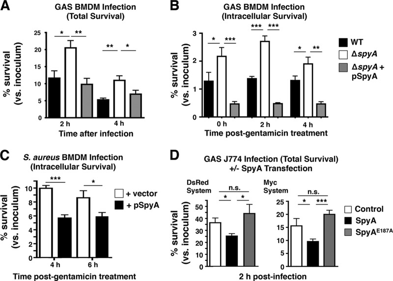
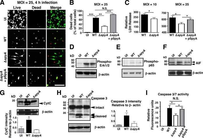
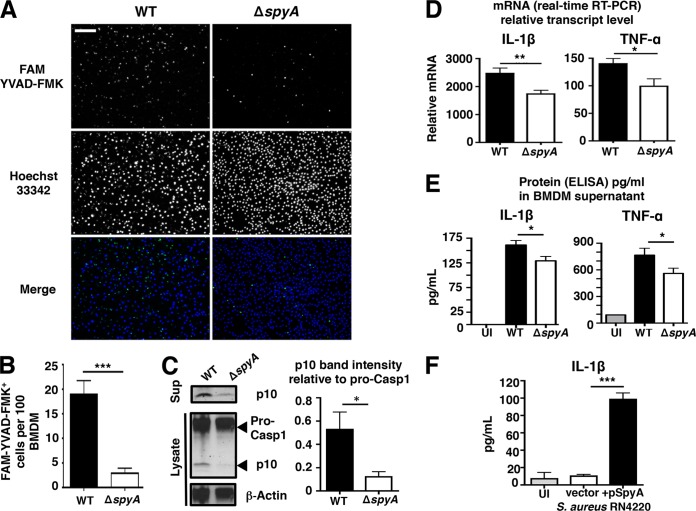
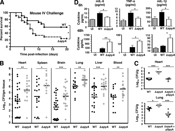
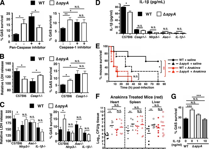
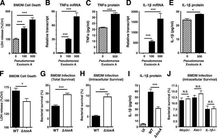
The M1T1 clone of group A Streptococcus (GAS) is associated with severe invasive infections, including necrotizing fasciitis and septicemia. During invasive M1T1 GAS disease, mutations in the covRS regulatory system led to upregulation of an ADP-ribosyltransferase, SpyA. Surprisingly, a GAS ΔspyA mutant was resistant to killing by macrophages and caused higher mortality with impaired bacterial clearance in a mouse intravenous challenge model. GAS expression of SpyA triggered macrophage cell death in association with caspase-1-dependent interleukin 1β (IL-1β) production, and differences between wild-type (WT) and ΔspyA GAS macrophage survival levels were lost in cells lacking caspase-1, NOD-like receptor protein 3 (NLRP3), apoptosis-associated speck-like protein (ASC), or pro-IL-1β. Similar in vitro findings were identified in macrophage studies performed with pseudomonal exotoxin A, another ADP-ribosylating toxin. Thus, SpyA triggers caspase-1-dependent inflammatory cell death in macrophages, revealing a toxin-triggered IL-1β-dependent innate immune response pathway critical in defense against invasive bacterial infection.

Importance: Group A Streptococcus (GAS) is a leading human pathogen capable of producing invasive infections even in healthy individuals. GAS bacteria produce a toxin called SpyA that modifies host proteins through a process called ADP ribosylation. We describe how macrophages, frontline defenders of the host innate immune system, respond to SpyA by undergoing a specialized form of cell death in which they are activated to release the proinflammatory cytokine molecule interleukin 1β (IL-1β). Release of IL-1β activates host immune cell clearance of GAS, as we demonstrated in tissue culture models of macrophage bacterial killing and in vivo mouse infectious-challenge experiments. Similar macrophage responses to a related toxin of Pseudomonas bacteria were also shown. Thus, macrophages recognize certain bacterial toxins to activate a protective immune response in the host.


Figures

FIG 1
FIG 1
SpyA-deficient GAS bacteria evade macrophage killing. To measure total and intracellular killing, BMDMs isolated from C57BL/6 mice were activated through overnight incubation in DMEM supplemented with 2% FBS. (A) Total killing was measured by recovering total CFU of cells infected with GAS (AP) at an MOI of ~10 after 2 and 4 h. (B) Intracellular killing was assessed by recovering CFU from cells infected with GAS for 30 min followed by 100 µg/ml of gentamicin treatment (1 h); levels of internalized bacteria were monitored over 2 and 4 h in serum-free media. (C) BMDMs were infected with S. aureus RN4220 at an MOI of ~5, and intracellular killing was monitored at 4 and 6 h. (D) Transfected J774 cells were infected with ΔspyA GAS for 2 h in 2% FBS media for a total killing assay. Data shown are representative of the results of multiple repeats. Error bar; SEM. *, P < 0.05; **, P < 0.01; ***, P < 0.001; n.s. = not significant; n = 3 (Student’s two-tailed unpaired t test or one-way ANOVA and Tukey’s multiple-comparison test).
FIG 2
FIG 2
SpyA promotes cell death and caspase-3 activation in a manner independent of MAPK and NF-κB signaling. (A) LIVE/DEAD staining illustrating BMDM viability after 4 h of GAS (AP) infection. Cells were infected at an MOI of ~25 or remained uninfected (UI). Scale bar, 100 µM. (B) Numbers of dead cells were counted from multiple field of views (n = 12). (C) Relative levels of LDH release (percentages compared to uninfected [UI] cells). ΔspyA-infected BMDMs exhibit reduced 2-h cell cytotoxicity at an MOI of 10 or 25. Infection with the SpyA-complemented strain restored LDH release to the WT level (n = 4). Error bars, SEM. *, P < 0.05; **, P < 0.01; ***, P < 0.001 (Student’s two-tailed unpaired t test). (D to H) Western blot analysis of whole BMDM lysates after 2 h of GAS infection. β-Actin was used as a loading control. (D) Phospho-ERK1/2 (42 or 44 kDa). (E) Phospho-p-p65 (65 kDa). (F) Apoptosis-inducing factor (AIF; 58 or 68 kDa). (G) Cytochrome c (15 kDa). (H) Caspase-3 (FL, full length [35 kDa]; CLVD, cleaved [17 or 19 kDa]). (I) Capsase-3 and caspace-7 (Caspace-3/7) activity assay. Error bars, SEM. *, P < 0.05; n = 4 (Student’s two-tailed unpaired t test).
FIG 3
FIG 3
SpyA stimulates a caspase-1-dependent inflammatory response and IL-1β production in macrophages. (A) BMDMs were infected with GAS (AP) at an MOI of ~10. At the end of the assay, cells were washed and stained for 1 h with FAM YVAD-FMK to visualize activated caspase-1 (green) and for 5 min with Hoechst 33342 to visualize DNA (blue) under an epifluorescent microscope. Scale bar, 100 µm. (B) Quantification of FMK YVAD-FMK-positive cells (per 100 cells) from multiple random fields of view (n = 10) per sample. (C) Western blot illustrating the abundance of pro-caspase-1 and its p10 subunit in GAS-infected BMDM cell lysates and caspase-1 p10 subunit released in the supernatants (Sup). Data represent densitometry quantifications illustrating average levels of active caspase-1 p10 expression in lysates and supernatants relative to pro-caspase 1 expression. Error bars, SEM. *, P < 0.05 (n = 3). Experiments were performed in duplicate using BMDMs isolated from four different C57BL/6 mice. (D and E) Real-time qPCR (D) and ELISA (E) quantification of IL-1β and TNF-α transcripts and proteins produced by BMDMs infected with GAS 2 h after total killing (n > 3). (F) IL-1β released by BMDMs after 4 h infection with S. aureus RN4220 expressing pSpyA or control vector (n = 4). Error bars, SEM. *, P < 0.05; **, P < 0.01; ***, P < 0.001 (Student’s two-tailed unpaired t test). UI, uninfected.
FIG 4
FIG 4
GAS SpyA expression is associated with increased bacterial clearance and reduced mortality in mice. CD1 mice were infected with 2 × 10⁵ to 4 × 10⁵ CFU of WT, ΔspyA, or ΔspyA plus pSpyA GAS (AP) via tail-vein injection. (A) Survival kinetics of infected mice monitored over 20 days (WT strain n = 20, ΔspyA strain n = 21). *, P = 0.02. Statistical significance was evaluated using the log rank test with a 95% confidence interval. (B) Bacteria recovered from organs or blood of GAS-infected mice 48 h postinfection for CFU enumeration to assess bacterial burden (n > 12). (C) CFU levels recovered from heart and lung of mice infected with ΔspyA plus pSpyA GAS (48 h) are significantly lower than those from ΔspyA mutant-infected mice (n = 6). (D) ELISA of proinflammatory cytokines from mouse blood serum isolate 6 h and 48 h postinfection (n = 5 to 9). UI = uninfected control, WT = wild-type GAS. Error bars, SEM. *, P < 0.05; **, P < 0.01; ***, P < 0.001 (Student’s two-tailed unpaired t test).
FIG 5
FIG 5
GAS SpyA activation of a caspase-1-dependent inflammasome response is required for bacterial clearance by cultured BMDMs and in vivo. (A) BMDMs were coincubated with GAS (AP) at an MOI of 20 plus 10 µM pan-caspase inhibitor zVAD-FMK (ZVAD) or 25 µM caspase-1 inhibitor Ac-YVAD-CHO (YVAD) for 2 h. Data shown are representative of the results of multiple experiments. (B) Relative levels of LDH release (percentages compared to levels in uninfected [UI] cells; left panel) and bacterial killing (right panel) by WT and Casp-1−/− BMDMs 2 h postinfection. (C) Relative levels of LDH release (left panel) and bacterial killing (right panel) 2 h postinfection (MOI = 5) by BMDMs isolated from wild-type (C57bl6), Nlrp3−/−, Asc−/−, and IL-1β−/− mice (n = 3). Data shown are representative of the results from two or more animals. (D) ELISA shows IL-1β secreted by BMDMs isolated from wild-type (C57bl6), Nlrp3−/−, Asc−/−, and IL-1β−/− mice after 2 h coincubation with GAS at an MOI of 20. Data shown are representative of the results from two animals. (E) CD1 mice were infected with 7 × 10⁵ CFU of WT or ΔspyA GAS (AP) via tail-vein injection (n = 7 per group). Immediately after infection, anakinra (IL-1 receptor antagonist) was subcutaneously injected at 100 mg/kg over a 12-h interval until the end of survival curve. WT plus saline solution versus ΔspyA plus saline solution, *, P = 0.04; WT plus saline solution versus WT plus anakinra. **, P = 0.0036; WT plus anakinra versus ΔspyA plus anakinra, P = 0.06; ΔspyA plus saline solution versus ΔspyA plus anakinra, P = 0.07 (log rank test with 95% confidence interval); N.S., not significant (P > 0.05). (F) GAS-infected mice (n = 8 to 10) were sacrificed 2 days postinfection; heart, spleen, and liver were harvested for CFU enumeration; no statistically significant differences were observed between WT and ΔspyA-infected animals that received anakinra treatment or for ΔspyA-infected animals with saline solution treatment. Closed symbols = WT; open symbols = ΔspyA. Black = control mice, red = anakinra-treated mice. Error bars, SEM. Statistical analysis: *, P < 0.05; N.S., not significant (P > 0.05) (Student’s two-tailed unpaired t test). (G) BMDMs in RPMI plus 2% FBS were treated with 1 or 10 ng/ml of recombinant mouse IL-1β (PeproTech, Rocky Hill, NJ) at the time of infection. Cells were washed and CFU recovered 2 h postinfection to assess bacterial killing. Error bars, SEM. *, P < 0.05; **, P = 0.01; ***, P < 0.001; NS, not significant; n = 3 (Student’s unpaired two-tailed t test).
FIG 6
FIG 6
Pseudomonas aeruginosa ADP-ribosyltransferase exotoxin A triggers an inflammasome-dependent macrophage response to enhance bacterial killing. BMDMs were seeded in 24 wells in 2% FBS media a day before infection. (A) LDH released by BMDMs treated with 100 ng or 500 ng of P. aeruginosa exotoxin A (PEA) for 18 h. (B to E) RNA and supernatants from BMDMs were isolated for real-time qPCR and ELISA to assess transcript and protein levels of TNF-α and IL-1β in BMDMs treated with PEA. (F) LDH assay of supernatants harvested from BMDMs 2 h after P. aeruginosa infection (n = 4). (G and H) Total and intracellular killing of P. aeruginosa infected for 30 min at an MOI of ~20, followed by a 1-h streptomycin treatment (150 µg/ml). Cells were washed and incubated in serum-free media for 2 h to establish intracellular killing. (I) ELISA of IL-1β produced by P. aeruginosa-infected BMDMs 2 h postinfection. (J) BMDMs from Nlrp3−/−, Asc−/−, and IL-1β−/− mice were isolated to assess intracellular killing of P. aeruginosa (2 h post-gentamicin treatment). Error bars, SEM. *, P < 0.05; **, P < 0.01; ***, P < 0.001; n = 3 (Student’s unpaired t test or one-way ANOVA followed by Dunnett’s test).

Similar articles

Cited by

References

    1. Carapetis JR, Steer AC, Mulholland EK, Weber M. 2005. The global burden of group A streptococcal diseases. Lancet Infect Dis 5:685–694. doi:10.1016/S1473-3099(05)70267-X. - DOI - PubMed
    1. Aziz RK, Kotb M. 2008. Rise and persistence of global M1T1 clone of Streptococcus pyogenes. Emerg Infect Dis 14:1511–1517. doi:10.3201/eid1410.071660. - DOI - PMC - PubMed
    1. Sumby P, Whitney AR, Graviss EA, DeLeo FR, Musser JM. 2006. Genome-wide analysis of group A streptococci reveals a mutation that modulates global phenotype and disease specificity. PLoS Pathog 2:e5. doi:10.1371/journal.ppat.0020005. - DOI - PMC - PubMed
    1. Cole JN, Barnett TC, Nizet V, Walker MJ. 2011. Molecular insight into invasive group A streptococcal disease. Nat Rev Microbiol 9:724–736. doi:10.1038/nrmicro2648. - DOI - PubMed
    1. Walker MJ, Hollands A, Sanderson-Smith ML, Cole JN, Kirk JK, Henningham A, McArthur JD, Dinkla K, Aziz RK, Kansal RG, Simpson AJ, Buchanan JT, Chhatwal GS, Kotb M, Nizet V. 2007. DNase Sda1 provides selection pressure for a switch to invasive group A streptococcal infection. Nat Med 13:981–985. doi:10.1038/nm1612. - DOI - PubMed

Publication types