. 2015 May;14(5):1373-84.
doi: 10.1074/mcp.M114.047381. Epub 2015 Mar 10.

Hinge-Region O-Glycosylation of Human Immunoglobulin G3 (IgG3)

Affiliations

Hinge-Region O-Glycosylation of Human Immunoglobulin G3 (IgG3)

Rosina Plomp et al. Mol Cell Proteomics. 2015 May.

Abstract

Immunoglobulin G (IgG) is one of the most abundant proteins present in human serum and a fundamental component of the immune system. IgG3 represents ∼8% of the total amount of IgG in human serum and stands out from the other IgG subclasses because of its elongated hinge region and enhanced effector functions. This study reports partial O-glycosylation of the IgG3 hinge region, observed with nanoLC-ESI-IT-MS(/MS) analysis after proteolytic digestion. The repeat regions within the IgG3 hinge were found to be in part O-glycosylated at the threonine in the triple repeat motif. Non-, mono- and disialylated core 1-type O-glycans were detected in various IgG3 samples, both poly- and monoclonal. NanoLC-ESI-IT-MS/MS with electron transfer dissociation fragmentation and CE-MS/MS with CID fragmentation were used to determine the site of IgG3 O-glycosylation. The O-glycosylation site was further confirmed by the recombinant production of mutant IgG3 in which potential O-glycosylation sites had been knocked out. For IgG3 samples from six donors we found similar O-glycan structures and site occupancies, whereas for the same samples the conserved N-glycosylation of the Fc CH2 domain showed considerable interindividual variation. The occupancy of each of the three O-glycosylation sites was found to be ∼10% in six serum-derived IgG3 samples and ∼13% in two monoclonal IgG3 allotypes.


Figures

Fig. 1.
Fig. 1.
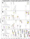
NanoLC-ESI-IT-CID spectra showing fragmentation of various trypsin-generated recombinant IgG3m(g) O-glycopeptides. Non-, mono- and disialylated core 1 type O-glycans (A–C) were seen attached to the peptide SCDTPPPCPR, as well as a disialylated O-glycan with an N-acetyllactosamine (N-acetylglucosamine + galactose) (D). These structures are partially based on literature since it is difficult to distinguish between different types of hexoses and N-acetylhexosamines with mass spectrometry. The triply charged peak at m/z 895.48 in panel D has a higher mass than the precursor mass, and thus likely originates from a contaminant. The MS1 precursor peak is shown for each fragmentation spectrum. Pep = peptide; yellow square = N-acetylgalactosamine, yellow circle = galactose; blue square = N-acetylglucosamine; purple diamond = N-acetylneuraminic acid.
Fig. 2.
Fig. 2.
A schematic overview of IgG3, which consists of two heavy chains (shown in dark gray, one variable and three conserved domains) and two light chains (light gray, one variable and one conserved domain). Black bars represent interchain disulfide bonds. An N-glycosylation site is present in domain CH2. Each IgG3 heavy chain contains three hinge repeat sequences with two or three partially occupied O-glycosylation sites. The peptide sequences of the major trypsin- and proteinase K-generated O-glycopeptides are shown.
Fig. 3.
Fig. 3.
(A) NanoLC-ESI-IT-MS/MS with electron transfer dissociation fragmentation of a sialidase- and galactosidase-treated tryptic glycopeptide of recombinant IgG3m(s). Peptide backbone fragmentation (c- and z-ions) and loss of the N-acetylhexosamine (HexNAc) or cysteine side chain (C) were observed. (B–C) CE-ESI-beam-type CID analysis was done on sialidase- and galactosidase-treated tryptic peptides of IgG3 derived from pooled plasma, and fragmentation spectra are shown for (B) the unoccupied tryptic hinge peptide SCDTPPPCPR and (C) the O-glycosylated version of the same peptide with a single N-acetylgalactosamine attached. The b- and y-ions are annotated. The MS1 precursor peak is shown on the right. The doubly charged peak at m/z 694.327 in panel C is likely a coeluting compound that appears in the spectrum because it was present within the precursor m/z window, and the peak at m/z 694.321 is the same compound after loss of water. The cysteine residues have been underlined to denote carbomidomethylation.
Fig. 4.
Fig. 4.
(A) Relative quantification of IgG3 O-glycosylation based on nanoLC-ESI-IT-MS analysis of tryptic glycopeptides from various IgG3 samples (IgG3 derived from pooled plasma, two monoclonal IgG3 allotypes and IgG3 purified from six donors (D1–6)). The signal intensities were normalized on the sum of all hinge-derived O-glycopeptides. The relative abundance and technical variation are based on LC-MS analyses of four distinct tryptic digests, each of them measured twice. The values given for glycopeptide NHS are expected to be significantly lower than the actual values because the triply charged compound overlapped with the doubly charged hinge peptide with a putative acetylation modification in all samples, leaving only the doubly charged peak for relative quantification. (B) An estimate of the total percentage of O-glycosylation was derived from relative quantification of tryptic (glyco)peptides that had been treated with exoglycosidases, trimming all O-glycans down to a single HexNAc. The averages and standard deviation are based on two LC-MS analyses of the same sample. (C) The number of N-acetylneuraminic acids per O-glycan was calculated from the same O-glycopeptide data listed under (A). A comprehensive list of values is available in supplemental Table S5.

References

    1. Stoop J. W., Zegers B. J., Sander P. C., Ballieux R. E. (1969) Serum immunoglobulin levels in healthy children and adults. Clin. Experiment. Immunol. 4, 101–112 - PMC - PubMed
    1. Morell A., Terry W. D., Waldmann T. A. (1970) Metabolic properties of IgG subclasses in man. J. Clin. Invest. 49, 673–680 - PMC - PubMed
    1. Vidarsson G., Dekkers G., Rispens T. (2014) IgG subclasses and allotypes: From structure to effector functions. Frontiers Immunol. 5, 520 - PMC - PubMed
    1. Roux K. H., Strelets L., Michaelsen T. E. (1997) Flexibility of human IgG subclasses. J. Immunol. 159, 3372–3382 - PubMed
    1. Dangl J. L., Wensel T. G., Morrison S. L., Stryer L., Herzenberg L. A., Oi V. T. (1988) Segmental flexibility and complement fixation of genetically engineered chimeric human, rabbit and mouse antibodies. EMBO J. 7, 1989–1994 - PMC - PubMed

Publication types

MeSH terms