. 2015 Feb 24:9:1193-208.
doi: 10.2147/DDDT.S72127. eCollection 2015.

Koenimbin, a natural dietary compound of Murraya koenigii (L) Spreng: inhibition of MCF7 breast cancer cells and targeting of derived MCF7 breast cancer stem cells (CD44(+)/CD24(-/low)): an in vitro study

Affiliations

Koenimbin, a natural dietary compound of Murraya koenigii (L) Spreng: inhibition of MCF7 breast cancer cells and targeting of derived MCF7 breast cancer stem cells (CD44(+)/CD24(-/low)): an in vitro study

Fatemeh Ahmadipour et al. Drug Des Devel Ther. .

Abstract

Background: Inhibition of breast cancer stem cells has been shown to be an effective therapeutic strategy for cancer prevention. The aims of this work were to evaluate the efficacy of koenimbin, isolated from Murraya koenigii (L) Spreng, in the inhibition of MCF7 breast cancer cells and to target MCF7 breast cancer stem cells through apoptosis in vitro.

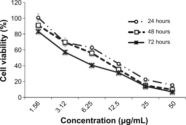
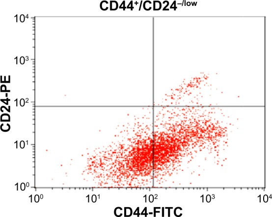
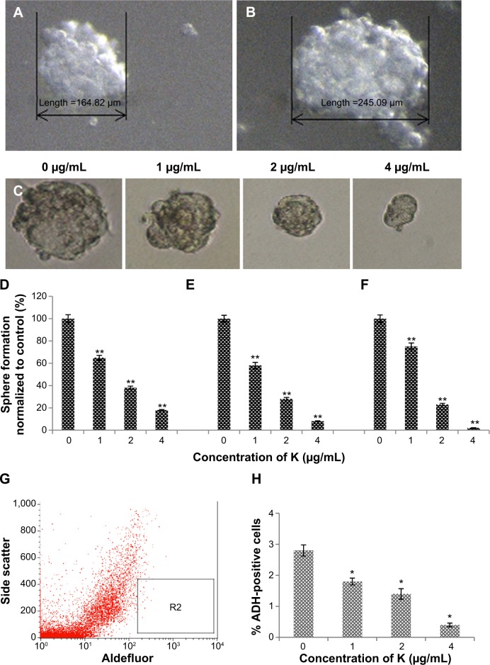
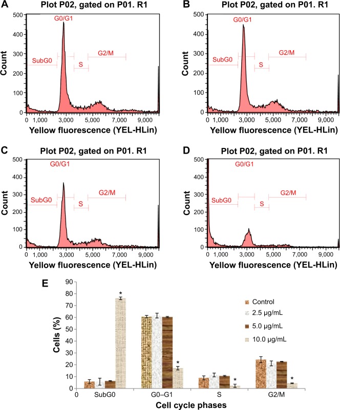
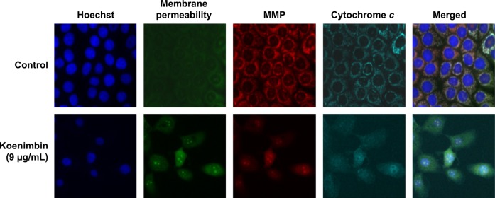
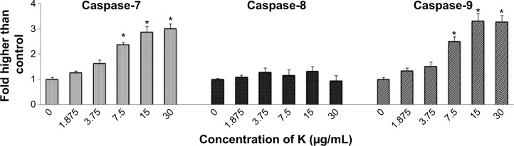
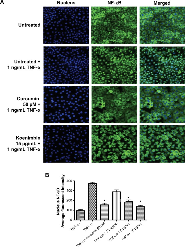
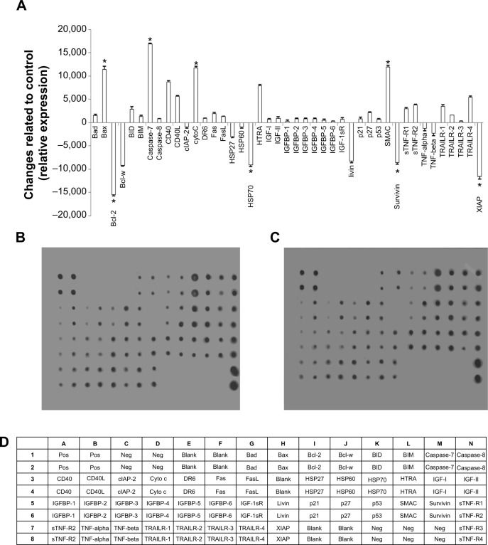
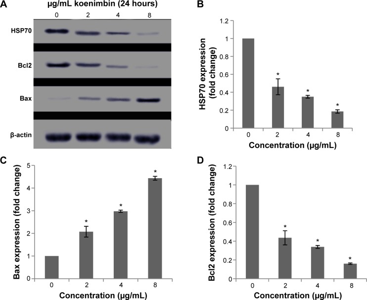
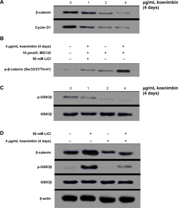
Methods: Koenimbin-induced cell viability was evaluated using the MTT (3-(4,5-dimethylthiazol-2-yl)-2,5-diphenyltetrazolium bromide) assay. Nuclear condensation, cell permeability, mitochondrial membrane potential, and cytochrome c release were observed using high-content screening. Cell cycle arrest was examined using flow cytometry, while human apoptosis proteome profiler assays were used to investigate the mechanism of apoptosis. Protein expression levels of Bax, Bcl2, and heat shock protein 70 were confirmed using Western blotting. Caspase-7, caspase-8, and caspase-9 levels were measured, and nuclear factor kappa B (NF-κB) activity was assessed using a high-content screening assay. Aldefluor™ and mammosphere formation assays were used to evaluate the effect of koenimbin on MCF7 breast cancer stem cells in vitro. The Wnt/β-catenin signaling pathway was investigated using Western blotting.

Results: Koenimbin-induced apoptosis in MCF7 cells was mediated by cell death-transducing signals regulating the mitochondrial membrane potential by downregulating Bcl2 and upregulating Bax, due to cytochrome c release from the mitochondria to the cytosol. Koenimbin induced significant (P<0.05) sub-G0 phase arrest in breast cancer cells. Cytochrome c release triggered caspase-9 activation, which then activated caspase-7, leading to apoptotic changes. This form of apoptosis is closely associated with the intrinsic pathway and inhibition of NF-κB translocation from the cytoplasm to the nucleus. Koenimbin significantly (P<0.05) decreased the aldehyde dehydrogenase-positive cell population in MCF7 cancer stem cells and significantly (P<0.01) decreased the size and number of MCF7 cancer stem cells in primary, secondary, and tertiary mammospheres in vitro. Koenimbin also significantly (P<0.05) downregulated the Wnt/β-catenin self-renewal pathway.

Conclusion: Koenimbin has potential for future chemoprevention studies, and may lead to the discovery of further cancer management strategies by reducing cancer resistance and recurrence and improving patient survival.

Keywords: MCF7 breast cancer stem cells; Murraya koenigii (L) Spreng; Wnt/β-catenin; glycogen synthase kinase 3β; koenimbin; nuclear factor kappa B.


Figures

Figure 1
Figure 1
MTT (3-(4,5-dimethylthiazol-2-yl)-2,5-diphenyltetrazolium bromide) assay. Growth curve for koenimbin-treated MCF7 cells at 24, 48, and 72 hours.
Figure 2
Figure 2
MCF7 cancer stem cells were identified by expression of CD44+ and low expression of CD24−/low in quadrant analysis (CD44+/CD24−/low). Each experiment was performed three times (n=3). Abbreviations: FITC, fluorescein isothiocyanate; PE, phycoerythrin.
Figure 3
Figure 3
Mammosphere formation and Aldefluor™ assay of MCF7 cancer stem cells. Notes: Size of mammospheres containing MCF7 cancer stem cells on day 5 (A) and day 7 (B). MCF7 cancer stem cells were cultured in mammosphere-forming conditions, and were incubated with koenimbin (0, 1, 2, and 4 μg/mL) for 7 days (magnification ×100) (C). Koenimbin reduced the size of the primary mammospheres. In the absence of drug, the second and third passages derived from koenimbin-treated primary mammospheres yielded smaller numbers of spheres in comparison with the control. The size of the mammospheres was estimated using V = (4/3)πR3. Koenimbin inhibits mammosphere formation and prevents self-renewal of (D) primary, (E) secondary, and (F) tertiary mammosphere-forming units. Data are shown as the mean ± standard deviation (n=3). **P<0.01 versus control. (G) Aldefluor assay of MCF7 cancer stem cells. Single cells obtained from cell cultures were incubated for 50 minutes at 37°C in Aldefluor assay buffer containing an ADH substrate, BODIPY-aminoacetaldehyde (1 μmol/L per 1×10⁶ cells). A cell population (R2) with high ADH activity was reported to enrich mammary stem/progenitor cells. (H) Inhibitory effect of koenimbin on ADH-positive cell populations. MCF7 cancer stem cells were treated with koenimbin 1, 2, or 4 μg/mL for 4 days and subjected to Aldefluor assay and flow cytometry analysis. Koenimbin decreased the percentage of ADH-positive cells. Data are shown as the mean ± standard deviation (n=3). *P<0.05 versus control; **P<0.01 versus control. Abbreviations: ADH, aldehyde dehydrogenase; K, koenimbin.
Figure 4
Figure 4
Cell cycle histograms from analyses of MCF7 cells treated with 0 (A), 2.5 (B), 5 (C), and 10 μg/mL (D) of koenimbin for 12 hours. (E) Summary of cell cycle progression for control and koenimbin-treated MCF7 cells. Notes: Data are shown as the mean ± standard deviation (n=3). *P<0.05 versus control.
Figure 5
Figure 5
Representative images of MCF7 cells treated with medium alone and koenimbin 9 μg/mL, and stained with Hoechst for nuclear, cytochrome c, membrane permeability, and MMP dyes, and cytochrome c dye. The images from each row are obtained from the same field of the same treatment sample (magnification 20×). Abbreviation: MMP, mitochondrial membrane potential.
Figure 6
Figure 6
Relative bioluminescence expression of caspase-7, caspase-8, and caspase-9 in MCF7 cells treated with koenimbin at various concentrations. Notes: The results are shown as the mean ± standard deviation of three independent experiments. Statistical significance is expressed as *P<0.05. Abbreviation: K, koenimbin.
Figure 7
Figure 7
(A) Photographs of intracellular targets in stained MCF7 cells treated with koenimbin for 3 hours and then stimulated for 30 minutes with TNF-α 1 ng/mL (NF-κB activation). (B) Representative bar chart indicating a significant decline in average fluorescent intensity of nuclei NF-κB, confirming that koenimbin inhibited TNF-α-induced translocation of NF-κB from the cytoplasm to the nucleus. Notes: Data are shown as the mean ± standard deviation (n=3). *P<0.05 versus control. Abbreviations: NF-κB, nuclear factor kappa B; TNF-α, tumor necrosis factor alpha.
Figure 8
Figure 8
Quantitative analysis of the human apoptosis proteome profiler array in koenimbin-induced MCF7 cells. MCF7 cells were lysed and protein arrays were performed. Cells were treated with koenimbin 9 μg/mL for 24 hours and total cell protein was extracted. Equal amounts (300 μg) of protein from each control and treated sample were used for the assay. Quantitative analysis of the arrays showed differences in the apoptotic markers. Notes: The graph shows the difference between treated cells as well as untreated control cells (A). Representative images of the apoptotic protein array are shown for the control (B), treated (C), and the exact protein name of each dot in the array (D). The results are shown as the mean ± standard deviation for three independent experiments. *Indicates a significant difference from control (P<0.05).
Figure 9
Figure 9
Western blot analysis of koenimbin in selected apoptotic signaling markers. Notes: The blot densities are expressed as fold of control (A). The HSP70 protein level was also downregulated, showing significant changes on treatment with koenimbin 4 and 8 μg/mL (B). The Bax (C) and Bcl2 (D) apoptotic markers were significantly elevated and reduced respectively, in a dose-dependent manner. The data are shown as the mean ± standard deviation (n=3). *P<0.05 versus control. Abbreviation: HSP, heat shock protein.
Figure 10
Figure 10
Western blot analysis of the Wnt/β-catenin self-renewal pathway in MCF7 cells treated with koenimbin. Koenimbin downregulated this pathway. Notes: Koenimbin decreased protein expression levels of β-catenin and cyclin D1 in MCF7 cells (A). Koenimbin increased the phospho-β-catenin Ser33/Ser37/Thr41, whereas LiCl suppressed phosphorylation through inactivation of GSK3β (B). Koenimbin decreased the expression level of p-GSK3β, while the protein expression of total GSK3β was unchanged (C). Koenimbin decreased β-catenin and LiCl-induced GSK3β phosphorylation, while LiCl elevated the protein expression level of β-catenin through GSK3β phosphorylation (D). Each experiment was performed three times (n=3). Abbreviation: GSK, glycogen synthase kinase.

Similar articles

Cited by

References

    1. Nakamura S, Nakashima S, Oda Y, et al. Alkaloids from Sri Lankan curry-leaf (Murraya koenigii) display melanogenesis inhibitory activity: structures of karapinchamines A and B. Bioorg Med Chem. 2013;21(5):1043–1049. - PubMed
    1. Ma Q, Tian J, Yang J, et al. Bioactive carbazole alkaloids from Murraya koenigii (L.) Spreng. Fitoterapia. 2013;87:1–6. - PubMed
    1. Arulselvan P, Subramanian S. Effect of Murraya koenigii leaf extract on carbohydrate metabolism studied in streptozotocin induced diabetic rats. Int J Biol Chem. 2007;1(1):21–28.
    1. Vinuthan M, Kumar VG, Narayanaswamy N, Veena T. Lipid lowering effect of aqueous leaves extract of Murraya koenigii (curry leaf) on alloxan-induced male diabetic rats. Pharmacogn Mag. 2007;3(10):112.
    1. Arulselvan P, Subramanian SP. Beneficial effects of Murraya koenigii leaves on antioxidant defense system and ultra structural changes of pancreatic β-cells in experimental diabetes in rats. Chem Biol Interact. 2007;165(2):155–164. - PubMed

Publication types

MeSH terms