. 2015 Feb 20:8:461-70.
doi: 10.2147/OTT.S78358. eCollection 2015.

Epigallocatechin-3-gallate suppresses cell proliferation and promotes apoptosis and autophagy in oral cancer SSC-4 cells

Affiliations

Epigallocatechin-3-gallate suppresses cell proliferation and promotes apoptosis and autophagy in oral cancer SSC-4 cells

Alexandra Iulia Irimie et al. Onco Targets Ther. .

Abstract

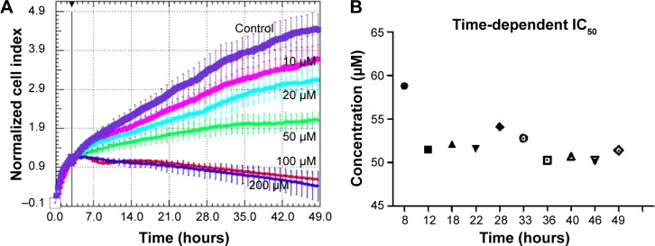
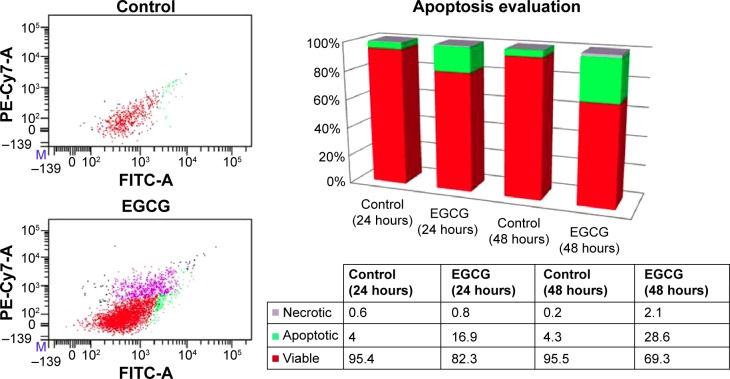
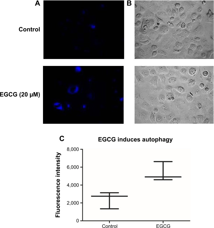
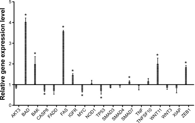
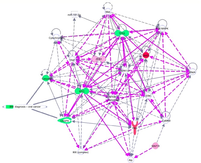
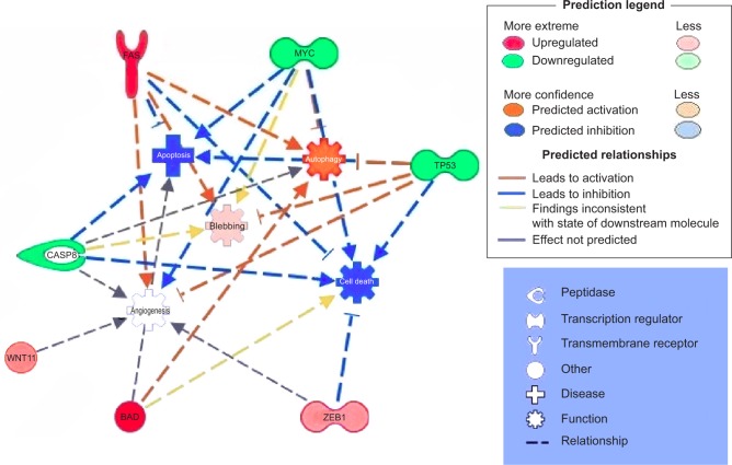
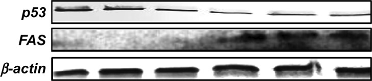
Epigallocatechin-3-gallate (EGCG) is the major bioactive component of green tea. Our experimental data indicated that EGCG treatment suppresses cell proliferation of SSC-4 human oral squamous cell carcinoma (OSCC), the effect being dose- and time-dependent. In parallel was observed the activation of apoptosis and autophagy, in response to EGCG exposure in SSC-4 cells. Treatment with EGCG activates the expression of the BAD, BAK, FAS, IGF1R, WNT11, and ZEB1 genes and inhibits CASP8, MYC, and TP53. All of these results suggest that EGCG has an excellent potential to become a therapeutic compound for patients with OSCC, by inducing tumor cell death via apoptosis and autophagy.

Keywords: gene expression; oral squamous carcinoma; time dependent cell proliferation.


Figures

Figure 1
Figure 1
Time-dependent cell proliferation evaluation for EGCG treatment at different EGCG doses (0, 10, 20, 50, 100, and 200 μM), in SSC-4 cells, using the xCELLigence system. Notes: (A) Data are displayed as the normalized cell index, over 49 hours. (B) Time-dependent IC50 evaluation using RTCA software. Abbreviations: EGCG, Epigallocatechin-3-gallate; IC50, half maximal inhibitory concentration; RTCA, real-time cell analysis.
Figure 2
Figure 2
Flow cytometry evaluation using annexin V FITC/PI, showing the effects of 20 μM EGCG treatment for 24 hours and 48 hours on SSC-4 cells. Notes: Viable cells are located in the lower left quadrant, and early apoptotic cells are positioned in the lower right quadrant of the two flow cytometric displays, while late apoptotic cells are located in the upper right quadrant, and necrotic cells are displayed in the upper left quadrant (24 hours). The image at the right presents the % apoptotic, necrotic, and viable cells, from total numbers of cells. Abbreviations: EGCG, epigallocatechin-3-gallate; FITC, fluorescein isothiocyanate; PI, propidium iodide.
Figure 3
Figure 3
EGCG-induced autophagy in SSC-4 cells, using fluorescence microscopy evaluation after MDC staining. Notes: (A) The control shows a low level of basal autophagy, indicated by blue “dot” staining of the autophagic vacuole, while MDC staining of SSC-4 cells treated with 20 μM EGCG led to an increase in fluorescence intensity and number of autophagic vacuoles compared with the untreated cells. (B) Bright-field microscopy. (C) Evaluation of the MDC fluorescence intensity, using a BioTek Microplate Reader. Abbreviations: EGCG, epigallocatechin-3-gallate; MDC, monodansylcadaverine.
Figure 4
Figure 4
Relative gene expression level showing the effect of treatment with a single dose of EGCG (20 μM), at 24 hours posttreatment. Abbreviation: EGCG, epigallocatechin-3-gallate.
Figure 5
Figure 5
Gene network, created using IPA® software, reflecting EGCG treatment. Notes: The overexpressed genes are labeled in red, while those downregulated are marked in green. Abbreviations: EGCG, epigallocatechin-3-gallate; IPA, Ingenuity® Pathway Analysis.
Figure 6
Figure 6
Putative model of multiple antitumoral mechanisms activated by EGCG, in SSC-4 human oral cancer cells. Notes: Overexpressed genes are labeled in red, and downregulated in green. Abbreviation: EGCG, epigallocatechin-3-gallate.
Figure 7
Figure 7
p53, FAS, and β-actin immunoblotting on SSC-4 cells, showing the effects of treatment with a single dose of EGCG (20 μM), at 48 hours posttreatment. Abbreviation: EGCG, epigallocatechin-3-gallate.

References

    1. Parris TZ, Aziz L, Kovács A, et al. Clinical relevance of breast cancer-related genes as potential biomarkers for oral squamous cell carcinoma. BMC Cancer. 2014;14:324. - PMC - PubMed
    1. Sand L, Jalouli J. Viruses and oral cancer. Is there a link? Microbes Infect. 2014;16(5):371–378. - PubMed
    1. Bhavana SM, Lakshmi CR. Oral oncoprevention by phytochemicals – a systematic review disclosing the therapeutic dilemma. Adv Pharm Bull. 2014;4(Suppl 1):S413–S420. - PMC - PubMed
    1. Lin LW, Lu YC, Wang CP, et al. Visfatin/pre-B cell colony-enhancing factor immunohistochemical overexpression in oral cancers. J Appl Biomed. 2014;12(4):255–226.
    1. González-Ramírez I, Soto-Reyes E, Sánchez-Pérez Y, Herrera LA, García-Cuellar C. Histones and long non-coding RNAs: the new insights of epigenetic deregulation involved in oral cancer. Oral Oncol. 2014;50(8):691–695. - PubMed