Review
. 2015:2015:859073.
doi: 10.1155/2015/859073. Epub 2015 Feb 1.

Intramyocardial hemorrhage: an enigma for cardiac MRI?

Affiliations
Review

Intramyocardial hemorrhage: an enigma for cardiac MRI?

Camilla Calvieri et al. Biomed Res Int. 2015.

Abstract

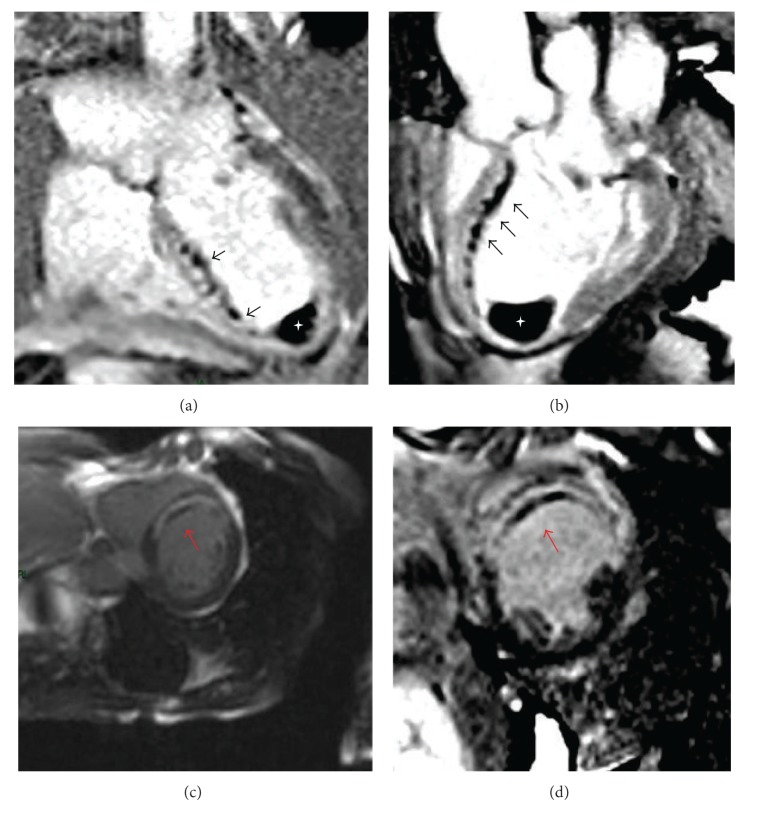
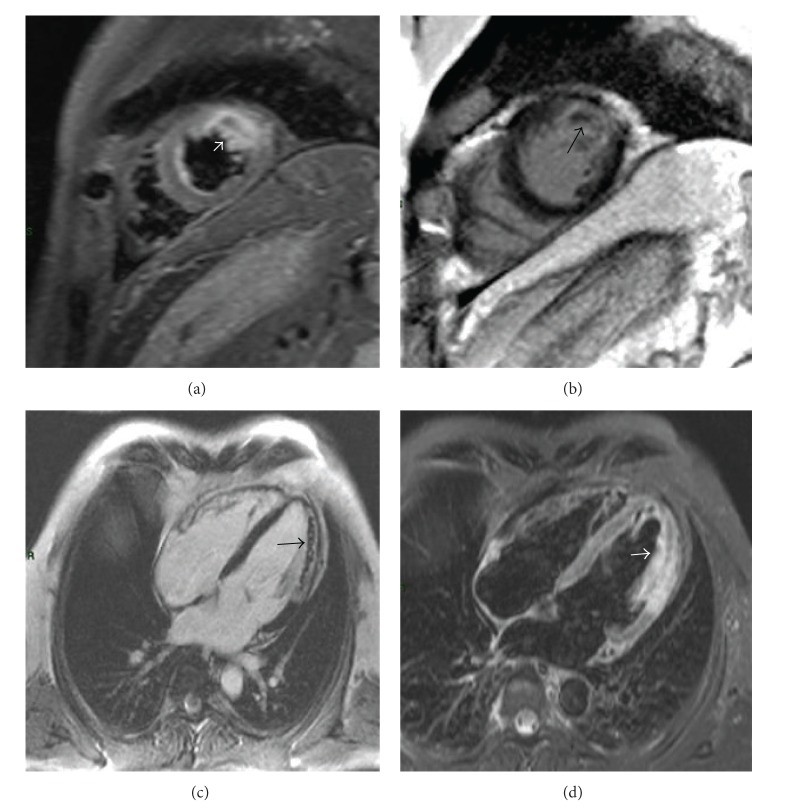
Cardiovascular magnetic resonance (CMR) is a useful noninvasive technique for determining the presence of microvascular obstruction (MVO) and intramyocardial hemorrhage (IMH), frequently occurring in patients after reperfused myocardial infarction (MI). MVO, or the so-called no-reflow phenomenon, is associated with adverse ventricular remodeling and a poor prognosis during follow-up. Similarly, IMH is considered a severe damage after revascularization by percutaneous primary coronary intervention (PPCI) or fibrinolysis, which represents a worse prognosis. However, the pathophysiology of IMH is not fully understood and imaging modalities might help to better understand that phenomenon. While, during the past decade, several studies examined the distribution patterns of late gadolinium enhancement with different CMR sequences, the standardized CMR protocol for assessment of IMH is not yet well established. The aim of this review is to evaluate the available literature on this issue, with particular regard to CMR sequences. New techniques, such as positron emission tomography/magnetic resonance imaging (PET/MRI), could be useful tools to explore molecular mechanisms of the myocardial infarction healing process.


Figures

Figure 1
Figure 1
Microvascular obstruction on EGE and LGE imaging. Cardiovascular magnetic resonance has been performed 5 days after PCI in a 54-year-old male patient with anterior ST-elevation myocardial infarction. On T1 early gadolinium enhancement (EGE) images ((a), (b)), the persistent microvascular damage appears dark (so-called “dark zones”) (black arrows) within late gadolinium enhancement area, representing the myocardial MVO on septal and anterior wall. White asterisk is the massive thrombus inside the ventricle apex ((a), (b)). T1 LGE short axis images of the same patient ((c), (d)) show transmural late gadolinium enhancement on the anteroseptal wall and a dark zone within this area indicating MVO (red arrows).
Figure 2
Figure 2
MVO and hemorrhagic areas in patient after reperfused anterolateral STEMI. ((a), (d)) In the anterolateral wall a large transmural hyperintense region and an hypointense core centrally located therein (white arrows) are present on the T2-short time inversion recovery (STIR) images in short axis and four chamber views showing edema on mid-apical anterolateral wall and an hypointense hemorrhagic area within the edema. ((b), (c)) Late gadolinium enhancement images show microvascular obstruction (black arrows), consistent with the hypointense area of haemorrhage, on mid-apical anterolateral wall.

References

    1. Rezkalla S. H., Kloner R. A. Coronary no-reflow phenomenon: from the experimental laboratory to the cardiac catheterization laboratory. Catheterization and Cardiovascular Interventions. 2008;72(7):950–957. doi: 10.1002/ccd.21715. - DOI - PubMed
    1. Niccoli G., Burzotta F., Galiuto L., Crea F. Myocardial no-reflow in humans. Journal of the American College of Cardiology. 2009;54(4):281–292. doi: 10.1016/j.jacc.2009.03.054. - DOI - PubMed
    1. Niccoli G., Cosentino N., Spaziani C., Fracassi F., Tarantini G., Crea F. No-reflow: incidence and detection in the cath-lab. Current Pharmaceutical Design. 2013;19(25):4564–4575. doi: 10.2174/1381612811319250005. - DOI - PubMed
    1. Bekkers S. C. A. M., Smulders M. W., Passos V. L., et al. Clinical implications of microvascular obstruction and intramyocardial haemorrhage in acute myocardial infarction using cardiovascular magnetic resonance imaging. European Radiology. 2010;20(11):2572–2578. doi: 10.1007/s00330-010-1849-9. - DOI - PMC - PubMed
    1. Lotan C. S., Bouchard A., Cranney G. B., Bishop S. P., Pohost G. M. Assessment of post reperfusion myocardial hemorrhage using proton NMR imaging at 1.5 T. Circulation. 1992;86(3):1018–1025. doi: 10.1161/01.CIR.86.3.1018. - DOI - PubMed

MeSH terms

LinkOut - more resources