. 2014:2014:635108.
doi: 10.1155/2014/635108. Epub 2014 Dec 23.

Human CD38hiCD138⁺ plasma cells can be generated in vitro from CD40-activated switched-memory B lymphocytes

Affiliations

Human CD38hiCD138⁺ plasma cells can be generated in vitro from CD40-activated switched-memory B lymphocytes

Rayelle Itoua Maïga et al. J Immunol Res. 2014.

Abstract

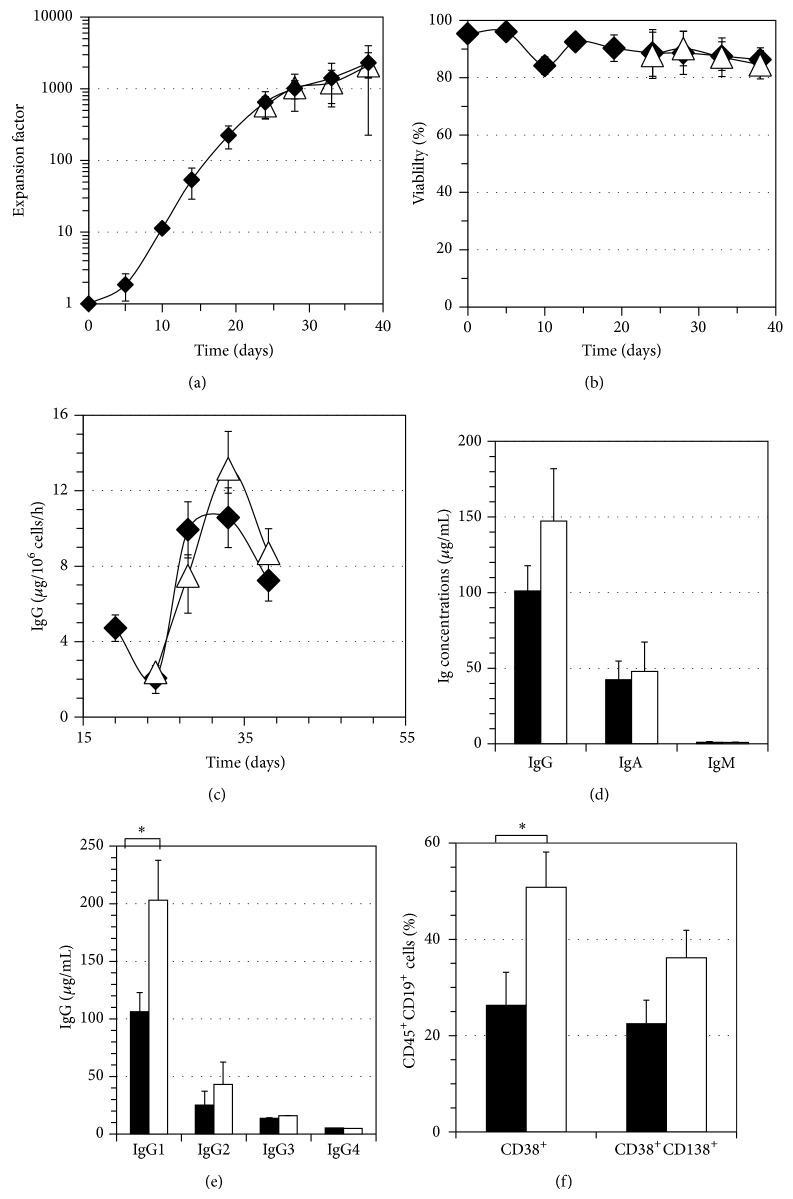
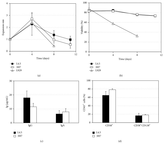
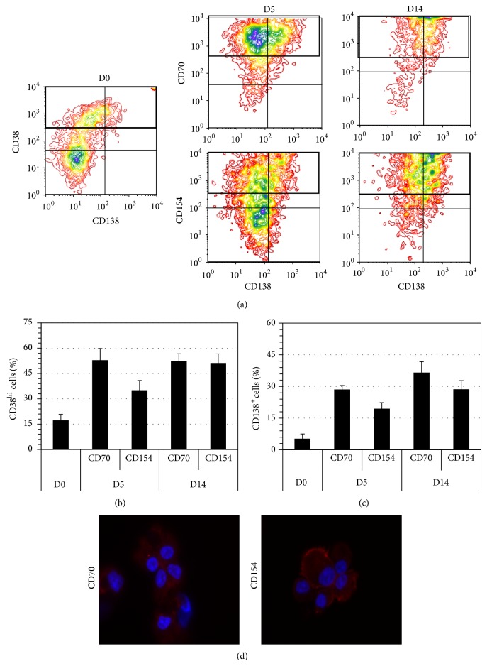
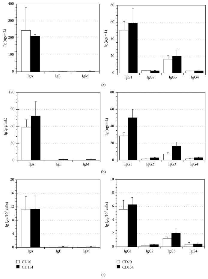
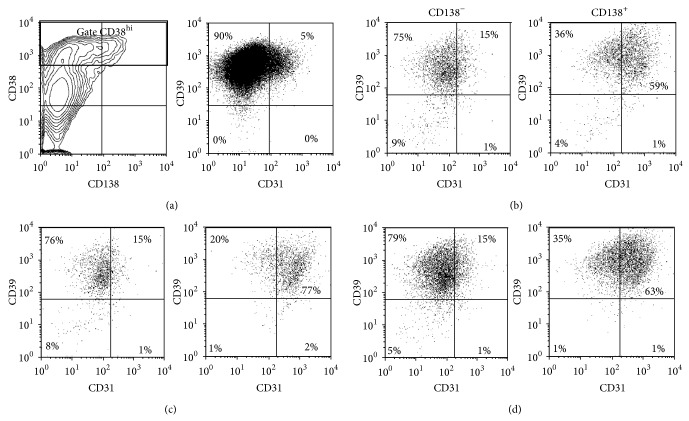
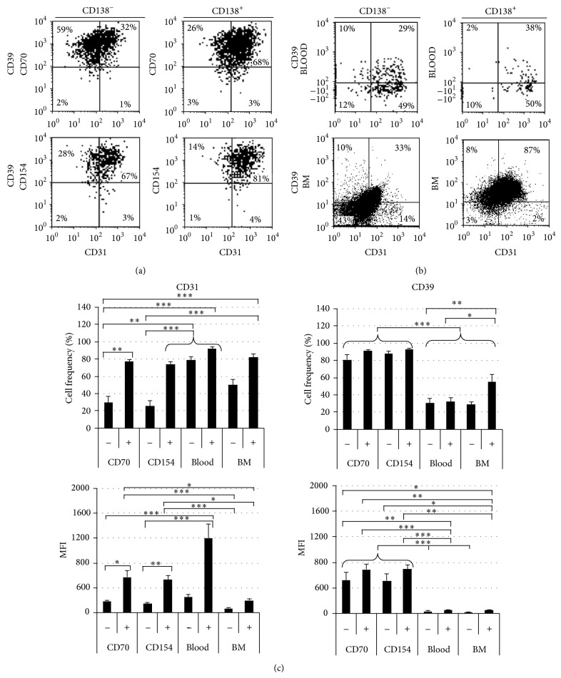
B lymphocyte differentiation into long-lived plasma cells is the keystone event for the production of long-term protective antibodies. CD40-CD154 and CD27-CD70 interactions are involved in human B lymphocyte differentiation into CD38(hi)CD138(+) cells in vivo as well as in vitro. In this study, we have compared these interactions in their capacity to drive switched-memory B lymphocytes differentiation into CD38(hi)CD138(+) plasma cells. The targeted B lymphocytes were isolated from human peripheral blood, expanded for 19 days, and then submitted to CD70 or CD154 interactions for 14 days. The expanded B lymphocytes were constitutively expressing CD39, whereas CD31's expression was noticed only following the in vitro differentiation step (day 5) and was exclusively present on the CD38(hi) cell population. Furthermore, the generated CD38(hi)CD138(+) cells showed a higher proportion of CD31(+) cells than the CD38(hi)CD138(-) cells. Besides, analyses done with human blood and bone marrow plasma cells showed that in vivo and de novo generated CD38(hi)CD138(+) cells have a similar CD31 expression profile but are distinct according to their reduced CD39 expression level. Overall, we have evidences that in vitro generated plasma cells are heterogeneous and appear as CD39(+) precursors to the ones present in bone marrow niches.


Figures

Figure 1
Figure 1
In vitro plasma cell generation and cytokine microenvironment. B lymphocytes were expanded for 19 days with IL-2, IL-4, and IL-10 at high interaction level with CD154. The same interleukin combination was used for the differentiation phase (filled symbols and bars) or the cells were cultured from day 19 with a combination of IL-6 and IL-10 (empty symbols and bars). The transition and differentiation phases lasted a total of 18 days with a low CD154 interaction. The results shown are the mean of 6 independent experiments. (a) Cell expansion, (b) cell viability, (c) IgG secretion rate in the differentiation environment, from D19 to D38, (d) IgG, A, and M secretion on day 33 supernatants, and (e) IgG subclasses secretion on day 33 supernatants. A significant difference was noticed in IgG1 secretion among the two interleukins combinations. Statistical analyses were done using the Bonferroni t-test and the P value was <0.05 (f) CD38 and CD38+CD138+ cells frequency. The IL-6-10 combination generated a larger CD38+ cell population on day 33, confirmed by a paired t-test, P value = 0.0188. All errors bars stand for SD and can be smaller than symbols.
Figure 2
Figure 2
Proliferation and viability following low CD70 and CD154 interaction. B lymphocytes were monitored following their stimulation with CD70 and CD154 as well as for their response to the mock L929 cell line using a low ratio (1 : 20) for all support cells. (a) Proliferation and (b) viability were measured on days 4, 8, and 11. The time laps between days 0 and 4 are the transition phase and differentiation phase was started on day 4 and lasted until day 11. (c) Secretion of IgG and IgA was measured by ELISA assay with the pooled supernatant harvested on days 6, 8, and 11. (d) At the end of the differentiation phase (day 11), B lymphocytes were analyzed using flow cytometry to evaluate the frequency of CD38+ cells and CD38+CD138+ cells. These results are representative of 5 independent experiments. All errors bars stand for SD and can be smaller than symbols.
Figure 3
Figure 3
Generation of plasma cells from switched-memory B lymphocytes. Monitoring of plasma cell generation in the differentiation phase was done by flow cytometry and all analyses were done on viable cell populations. The results are presented as the mean ± S.E.M. of 6 independent samples: (a) CD38 and CD138 expression profiles on D0, 5 and 14. D0 being the day that the expanded memory B cells were thawed and were put in culture with CD70+ or CD154+ cell lines. (b) CD38hi cells frequency. The comparison of CD38hi cells frequency at D5 was determined using a Mann-Whitney test, P value = 0.0260. For the monitoring of CD138+ cells frequency (c), the Kruskal-Wallis test was used and no significant difference was observed. (d) Generated plasma cells morphology at D14 shown by fluorescence microscopy (100x immersion oil objective). Blue: nucleus; red: actin. Representative cells are shown.
Figure 4
Figure 4
In vitro generated plasma cells secrete high level of IgA and IgG. Immunoglobulin secretion in supernatants was measured by the Bio-Plex human isotyping kit. The cumulated Ig concentration was determined on (a) day 5 (D5) after the transition phase and (b) day 14 (D14) at the end of differentiation. (c) Relative secretion was determined on D14 by normalizing immunoglobulin concentrations with total cell count on day 5, corresponding to the plateau in proliferation. The results are presented as the mean ± S.E.M. of 6 independent samples. No differences were observed between CD70 and CD154 conditions.
Figure 5
Figure 5
High frequency of CD39+ cells among the expanded B lymphocyte population. Viable cells were analyzed for CD31 and CD39 expression before their transfer into differentiation conditions (D0). (a) Profiles of total viable cells according to CD38 and CD138 (left) and to CD31 and CD39 (right) are shown. The CD38hiCD138+/− cells represented 22 ± 3% of the cell population in the presence of CD154 interaction (b) and 25 ± 6% in the presence of CD70 (c). They represented 14 ± 2% of the population when being in the simultaneous presence of both interactions (d). These cells were further analyzed for their expression of CD31 and CD39 according to their expression of CD138 as indicated in (b, c, and d). These results are representative of 11 and 10 independent experiments for the stimulation with L4.5 cells (b) or 3H7 cells (c), respectively. The results in (d) are representative of three independent experiments.
Figure 6
Figure 6
CD31 as an in vivo and in vitro generated plasma cell marker. Analyses were done on cells submitted to 14 days in differentiation conditions. (a) D14 viable CD38hi cells were analyzed based on their CD31 and CD39 expression. Plots are representative of 10 independent experiments for the CD70 interaction and 13 for the CD154 interaction. (b) Bone marrow and peripheral blood plasma cells (viable CD45+CD38hi). Plots are representative of 10 and 14 independent experiments for bone marrows and for peripheral blood, respectively. (c) Comparison of CD31 and CD39 expression and frequency on generated (CD70 and CD154) and in vivo (PB and BM) plasma cells. CD138 (−) and CD138+ (+) plasma cells expression profiles were compared using the Unpaired t- or the Mann-Whitney test (*indicating a significant difference with P values < 0.03). The expression profile of CD138 (−) cells from different origins was compared (**indicating significant differences with P values < 0.02) as well as CD138+ (+) plasma cells (***indicating significant differences with P values < 0.006) using the Kruskal-Wallis test.

Similar articles

Cited by

References

    1. Oracki S. A., Walker J. A., Hibbs M. L., Corcoran L. M., Tarlinton D. M. Plasma cell development and survival. Immunological Reviews. 2010;237(1):140–159. doi: 10.1111/j.1600-065X.2010.00940.x. - DOI - PubMed
    1. Shapiro-Shelef M., Calame K. C. Regulation of plasma-cell development. Nature Reviews Immunology. 2005;5(3):230–242. doi: 10.1038/nri1572. - DOI - PubMed
    1. Tarlinton D., Radbruch A., Hiepe F., Dörner T. Plasma cell differentiation and survival. Current Opinion in Immunology. 2008;20(2):162–169. doi: 10.1016/j.coi.2008.03.016. - DOI - PubMed
    1. Lacotte S., Decossas M., Le Coz C., Brun S., Muller S., Dumortier H. Early differentiated CD138high MHCII+ IgG+ plasma cells express CXCR3 and localize into inflamed kidneys of lupus mice. PLoS ONE. 2013;8(3) doi: 10.1371/journal.pone.0058140.e58140 - DOI - PMC - PubMed
    1. Kawano M. M., Mihara K., Huang N., Tsujimoto T., Kuramoto A. Differentiation of early plasma cells on bone marrow stromal cells requires interleukin-6 for escaping from apoptosis. Blood. 1995;85(2):487–494. - PubMed